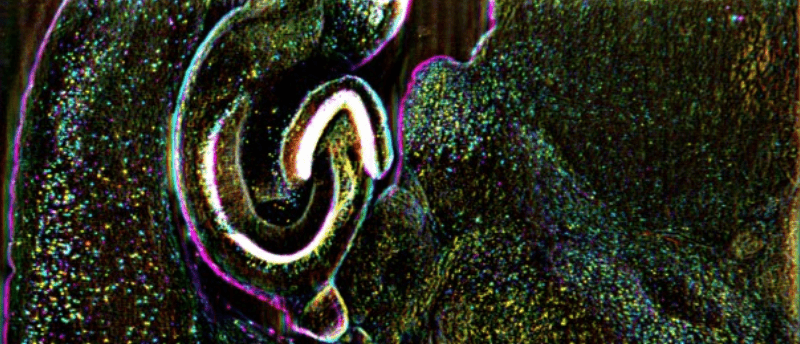
Oblique plane microscopy captures cell dynamics of whole organisms

Researchers developed a large-scale, dynamic imaging technique using mesoscopic oblique plane microscopy, which can capture 3D images of entire organisms and maintain cellular resolution.
A new microscopy technique can obtain dynamic 3D images of entire zebrafish larvae, enabling scientists to view how cells interact in their natural state. This technique, developed by researchers at Johns Hopkins University (MD, USA), could result in new insights used to develop treatments for diseases, among other applications.
“Unraveling underlying cellular structures and their interaction is fundamentally important to understanding life,” said Ji Yi, senior author of the study. “However, the limitations of light diffraction make it difficult to image with 3D cellular resolution over large areas of several millimetres.”
To capture 4D biological dynamics at cellular resolution, most microscopy techniques need to make a trade-off between the field of view, depth resolution and imaging speed. The researchers of this study circumvent this and maintain cellular resolution in all three dimensions using this mesoscopic oblique plane microscopy method.
 Expansion revealing microscopy exposes unseen cellular nanostructures
A technique called expansion revealing microscopy separates molecules in cells before fluorescence labeling, uncovering unseen nanostructures for the first time.
Oblique plane microscopy is a type of light-sheet microscopy that uses a sheet of laser light to illuminate a thin sample slice labeled with fluorescent markers, unlike a confocal microscope, which focuses a small beam at a sample. They reported that oblique plane microscopy captures up to three times more resolvable image points within an imaged 3D volume compared to other microscopy systems.
“Looking at biological systems in their larger context – also known as mesoscopic scale – provides holistic information for complex biological systems such as a complete neural circuit in mouse brain, 3D-tissue culture or entire zebrafish larvae,” explained Yi.
From microscopy to smartphone cameras, it is challenging to use a single set of optics to obtain an image that has a high resolution and large-scene coverage. To overcome this, smartphones often use more than one camera; however, this is not usually a feasible option for microscopy.
“Instead of adding another camera set, we used an optical component known as a transmission grating to create a diffractive light sheet,” said Yi. “This improves depth sectioning and resolution while using a low magnification lens. The result is the ability to perform mesoscopic-scale imaging over a field of view that is several millimeters wide while still being able to resolve individual cells in 3D.”
 Democratizing microscopy and diagnostics and developing digital holography
Aydogan Ozcan discusses his work in the democratization of microscopy and detection technologies, revealing the latest advances in digital holography.
To demonstrate the abilities of this mesoscopic oblique plane microscopy technique, the researchers imaged living zebrafish larvae, which were 3–4 mm long, and mouse brain slices containing living cells. Both of these expressed fluorescence proteins that labeled specific cell types including neurons and blood cells. The microscopy technique successfully imaged a 3D cellular structure with a single scan and achieved a field of view measuring up to 5.4 x 3.3 mm with a resolution of 2.5 x 3 x 6 microns.
The researchers captured a whole-body volumetric recording of the neural activity in the zebrafish larvae at a 2 Hz volume rate, meaning that neural circuits over the entire CNS can be studied in vertebrates with this technique. Additionally, whole-body volumetric recordings of blood flow dynamics were captured at 5 Hz in 3D cellular resolution. This allows single-cell tracking within a 3D circulation system for the first time.
This study lays the groundwork for further progress in developing biological imaging that is faster, with a larger field of view and deeper in samples. Next, the researchers are working to improve light collection efficiency so they can increase imaging speed. They also hope to incorporate multiphoton imaging to achieve better depth, which is another challenge in optical imaging.
You can find more information on the microscope setup in the paper.


