Radiotracer illuminates fructose metabolism

Researchers have developed a radiotracer that can detect fructose metabolism, paving the way for a clinically viable tool for the earlier detection of multiple conditions.
Fructolysis, the action of metabolizing fructose, has been associated with neurodegenerative conditions, such as Alzheimer’s disease, and cardiac disorders. Additionally, it has been hypothesized that the switch from glucose to fructose energy consumption could be a driver of cancer, particularly in solid tumors.
Positron emission tomography (PET) imaging is a key diagnostic tool for the detection of these conditions and currently glucose is the go-to sugar for PET imaging, as inflammatory and cancerous tissues consume higher levels of glucose than healthy tissues.
However, a conundrum arises when diagnosing cardiac and neural conditions as healthy heart and brain tissue naturally utilize high levels of glucose, which can mask signs of disease in these organs. This has led to researchers looking for alternatives to glucose. Fructose is a prime candidate as it is not usually used as a fuel within healthy brain and heart tissue.
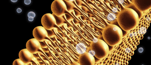
 Ferroptosis: two-tail fats discovered as the instigators
Researchers have uncovered that phospholipids with two polyunsaturated fatty acyl tails play a crucial role in driving ferroptosis.
In this study, scientists from the University of Ottawa (Ontario, Canada) sought to noninvasively map fructose metabolism to overcome the limitations of glucose imaging.
To start off, the team closely examined the catalytic mechanism of aldolase, the enzyme for which fructose-1-phosphate is a substrate. This enabled them to create a radiodeoxyfluorinated fructose analog, termed [18F]4-FDF.
The team then worked on validating and testing their radiotracer in both cell and animal models alongside comparing it to commonly used PET radiotracers, such as the glucose analog [18F]FDG and [18F]6-FDF.
Of note, the team demonstrated that [18F]4-FDF performed better than its glucose counterpart as it did not accumulate in bones and showed a low uptake in healthy brain and heart tissue. Using [18F]4-FDF PET/CT, the team were able to sensitively map both neuro- and cardio-inflammatory responses to fructose.
“For the first time, we can see where fructose, a common dietary sugar, is used in the body. Outside of the kidneys and the liver, fructose metabolism in any other organs may point to a sinister problem including cancer and inflammation,” explained study author Adam Shuhendler.
The team hope their findings will enable the visualization of increased fructose in diseased tissue, with the ultimate aim of creating a fructose-based diagnostic imaging biomarker.


