Bioprinting and bioinks: the latest innovations in building synthetic biological structures
Bioprinting is a bioengineering technique that utilizes the principles of 3D printing to build 3D tissue-like structures out of bioinks mixed with living cells. First making an appearance in 1988, bioprinting has since evolved, leading to the advent of new technologies and techniques that have broadened bioprinting’s applications from tissue engineering to drug discovery.
The bioprinting technique that’s expediting functional tissue creation
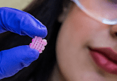
Researchers at Penn State University (PA, USA) have tried to overcome the limitations of previous bioprinting technologies – which are unable to print tissues with high cell density at scale – by developing a novel technique that uses spheroids to create complex tissues. Bioprinting tissues with a cell density that mimics that of the human body is difficult but essential to developing tissue that’s functional. That’s why the team focused on spheroids, high-density clusters of cells that more closely mimic the cell density seen in the human body.
In addition to tackling this density issue, the team also faced issues with scalability and slow processing. To overcome these limitations, they developed a new technique called High-throughput Integrated Tissue Fabrication System for Bioprinting (HITS-Bio), which uses a digitally controlled arrangement of multiple nozzles moving in three dimensions to manipulate several spheroids at once. Using this technique, the team can bioprint spheroids 10-times faster than existing techniques while maintaining over 90% cell viability. The method was successfully utilized to fabricate cartilage tissue as well as repair wounds in a rat model.
“This technique is a significant advancement in rapid bioprinting of spheroids,” commented Ibrahim Ozbolat, lead author of the study. “It enables the bioprinting of tissues in a high-throughput manner at a speed much faster than existing techniques with high cell viability.”
Read more here | Press release
 Bioprinting the brain: 3D models for Alzheimer’s research
Marimélia Porcionatto’s work focuses on biofabrication of 3D systems for studying the central nervous system, with an emphasis on neurodegenerative diseases and neurodevelopment. We caught up with her to discuss the 3D bioprinted models she’s developing!
The bioprinting technique that’s facilitating shape-morphing human heart tissues
University of Galway (Ireland) researchers, led by Ankita Pramanick and Andrew Daly, have developed a method of bioprinting tissues that change shape because of cell-generated forces, more closely mimicking the function of biological tissues during development. Focusing specifically on building functional heart tissue, the research team wanted to ensure their tissue was able to contract with the force measured in human adults. They recognized that dynamic cell-shape changes during natural embryonic development are an essential component of creating mature heart tissue, so they developed a new technique.
“Our work introduces a novel platform, using embedded bioprinting to bioprint tissues that undergo programmable and predictable 4D shape-morphing driven by cell-generated forces. Using this new process, we found that shape-morphing improved the structural and functional maturity of bioprinted heart tissues,” concluded Pramanick.
This was achieved by modifying factors such as initial print geometry and bioink stiffness. They found that this technique delivered heart tissue that contracted with greater strength, with greater similarity to the human heart.
Read more here | Press release
 Recreating the tumor microenvironment with 3D bioprinting technology
Cancer models retaining original tumor characteristics can be developed through 3D bioprinting, allowing for rapid evaluation of drug responses.
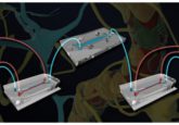
Bioprinting builds an accurate blood–brain barrier (BBB) model
To investigate the role of neuroinflammation – caused by interactions between neural cells and cerebral blood vessels – in neurodegenerative diseases, researchers at Pohang University of Science and Technology (POSTECH) and Seoul National University Hospital (both Republic of Korea) developed a 3D-bioprinted model of the BBB, which plays an important role in regulating neural cell–cerebral blood vessel interactions.
Although previous BBB models have been built, they lack the complex 3D structure of cerebral blood vessels. To overcome this limitation, the current research team developed a cerebrovascular-specific bioink using decellularized extracellular matrix (CBVdECM) derived from the brain and blood vessels of pigs. Additionally, they utilized 3D bioprinting technology to construct a tubular vascular model that precisely replicated the anatomical structure and function of the human BBB. The CBVdECM bioink also featured human brain microvascular endothelial cells and human brain vascular pericytes, which self-assembled into inner and outer vascular walls that closely resembled the architecture of biological blood vessels. What’s more, when exposed to inflammatory substances, such as TNF-α and IL-1β, the model reacted appropriately, modeling neuroinflammatory mechanisms that provide insights into BBB dysfunction and inflammation in neurodegenerative diseases.
“This study provides a crucial platform for investigating the pathological mechanisms of neuroinflammation and developing novel therapeutic strategies,” remarked co-senior author Jinah Jang (POSTECH). “We aim to integrate additional cell types, such as glial cells, neurons, and immune cells, to refine methods for quantifying inflammatory responses and permeability, while also expanding to patient-specific disease models.”
Read more here | Press release

BIO INX (East Flanders, Belgium), an innovator in materials for 3D bioprinting, has launched BIORES INX. This gelatin methacrylamide-based resin provides improved biocompatibility, meeting ISO 10993-5 standards, while offering simple processing and compatibility with digital light-processing bioprinting platforms.
Wound-healing nanocellulose bioink derived from kombucha
Researchers led by Insup Noh from Seoul National University of Science and Technology (Republic of Korea) have developed a nanocellulose bioink derived from Kombucha SCOBY (symbiotic culture of bacteria and yeast). The bioink offers a sustainable alternative to conventional bioink materials; it can also be loaded into a handheld ‘Biowork’ biopen, a device developed by the same team that allows for the precise application of bioink to damaged tissue areas.
The microorganisms in Kombucha SCOBY produce cellulose, which is biodegradable and compatible with cells. However, to utilize this cellulose for bioprinting purposes, its rheological and mechanical properties need to be modified. The current team partially hydrolyzed the cellulose with acetic acid, breaking glucose bonds and deconstructing its entangled structure to improve extrusion. This, in turn, caused the cellulose to be too weak to maintain its structure once printed. The team reinforced the cellulose with chitosan and kaolin nanoparticles, which electrostatically interact with cellulose to form a stable hydrogel suitable for 3D printing. The cellulose-derived hydrogel could then be loaded into a biopen along with living cells and mixed to form a homogenous bioink that could be applied directly into damaged tissue.
This technology allows for a quick and easy one-step process where the drug and hydrogel are mixed and immediately applied on-site to injured areas of different shapes,” concluded Noh.
Read more here | Press release
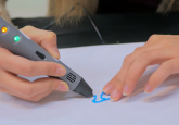
 A wound-healing bioink accelerates skin repair
Researchers have developed a bioactive hydrogel ink containing macrophage-derived extracellular vesicles that enhances cutaneous wound healing and can be administered in situ using a 3D-printing pen.
Bioprinting for bone and soft tissue repair
Hefei Institutes of Physical Science (Chinese Academy of Sciences; China) researchers, led by Wang Junfeng, have developed novel 3D bioprinting materials for bone and soft tissue repair. The team sought to address the challenges associated with building effective scaffolds for bone and soft tissue repair by turning their attention to combining known biomaterials like boron-based glass (BBG), polycaprolactone (PCL) and sodium alginate, which have been individually used previously for different bioprinting endeavors.
They proceeded to develop BBG/PCL composites with varying BBG contents using selective laser sintering technology and applied these composites to bone repair studies. Analysis of these composites’ pore geometry, porosity, mechanical strength, degradation behavior and cell compatibility revealed that materials with 20% BBG were optimal for supporting bone healing and regeneration.
For soft tissue repair, the team developed a BBG/sodium alginate bioink with high-precision printing capabilities, improved structure and enhanced biocompatibility. This bioink encouraged cell adhesion and promoted the expression of soft tissue repair-associated genes and proteins.
“Our work will significantly enhance the potential of bioactive glass in 3D bioprinting materials, providing a crucial research foundation for developing novel bio-based 3D printing materials,” commented Kun Ma.