Lights, stimulate, measure!

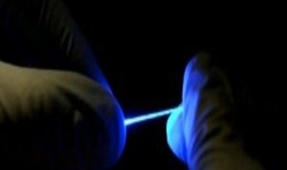
Stretchable implants record electrical signals in the spinal cords of freely moving mice, paving the way for advanced prosthetics that patients can control with spinal cord stimulation.

Credit: Chi (Alice) Lu and Seongjun Park
When building a house, discovering faulty wiring is not a big crisis; electricians can easily fix the problem. Addressing an analogous problem in our body, however, is not as simple. Injury to the spinal cord often results in paralysis, and our limited understanding of the spinal cord’s role in limb-function has yielded few therapeutic options.
Polina Anikeeva and her team at Massachusetts Institute of Technology develop neural probes for studying and manipulating the nervous system. In their recent work, described in the journal Science Advances, they engineered new conductive implants to stimulate and measure electrical activity in the spinal cord of mice. Data from these implants may help understand and manipulate spinal cord function, possibly facilitating recovery after injury.
Developing implants for the spinal cord was especially challenging, according to Seongjun Park, first author of the study. Spines bend and stretch during day-to-day movement, so the implant material had to be flexible to withstand large strains. It needed to be composed of soft materials to avoid damaging the spinal cord tissue, and be conductive so the researchers could use it to measure electric signals. “It is usually very hard to find a soft material with good conductivity,” Park explained.
By breaking down the probe preparation process into two steps, the team finally overcame these problems. First, they produced thin thread-like fibers from a transparent rubber-like material, elastomer, using a thermal drawing method. Then, they coated the elastomer fiber with a thin mesh of silver nanowires to make it conductive.
After verifying the mechanical, optical, and electrical properties of their elastomer implant, the team tested its biocompatibility by surgically inserting it into the spinal cords of healthy mice. Using the probe, they successfully recorded electrical signals. Next, they implanted the probe into mice with genetically modified neurons that can be triggered by blue light to fire an impulse. With this system, Park and his team demonstrated that they could optically control hind-limb movement in mice.
“This combination of optical, electrical, and mechanical properties in a single implantable probe poses as a significant step in electrophysiological activity recording and stimulation,” said Yue (Jessica) Wang, a postdoctoral researcher at Stanford University, an expert in stretchable polymers who was not involved in the study.
In the future, the team plans to collaborate with neuroscientists, physicians, and engineers towards the goal of implementing these probes in prosthetics to enable people to control new limbs via brain and spinal cord stimulation.


