Maturation of stem-cell-derived cardiomyocytes monitored with cyborg nanosensor
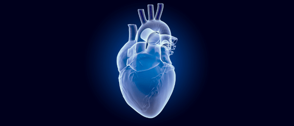
Novel tissue-embedded nanosensor shows that co-culturing stem-cell-derived cardiomyocytes with endothelial cells leads to more functionally mature cardiomyocytes.
Heart muscle cells, called cardiomyocytes, are responsible for heart contractions through synchronized electrical signals and cannot regenerate themselves following injury. Stem-cell-derived cardiomyocytes have shown promise in repairing and regenerating these heart muscle cells; however, further research is needed to ensure implanted cardiomyocyte cells integrate into the surrounding tissue effectively.
To study the functional development and maturation of cardiomyocytes, researchers at the Harvard John A. Paulson School of Engineering and Applied Sciences (MA, USA) developed a tissue-embedded nanoelectronic device that can monitor this continuously on a single-cell level.
The nanoelectronic device is flexible, stretchable and integrates into living cells to create ‘cyborgs’. “These mesh-like nanoelectronics, designed to stretch and move with growing tissue, can continuously capture long-term activity within individual stem-cell derived cardiomyocytes of interest,” explained Jia Liu, the co-senior author of the paper. “We were inspired by the way neural tubes fold during development, stretching as cells migrate and take shape into tissue volume.”
Liu’s research group has previously shown that these types of flexible nanoelectronic cyborgs can be safely implanted into living mice and do not disrupt the function of nearby cells.
 Introducing organoid intelligence: human brain cells that power biocomputers
I know you’ve heard of artificial intelligence, but what about organoid intelligence? Researchers from multiple disciplines are working together to create biocomputers powered by 3D cultures of human brain cells.
In this study, Liu collaborated with a research group at the Harvard Stem Cell Institute (MA, USA) led by Richard Lee to monitor the electrical activity of stem-cell-derived cardiomyocytes using their nanoelectronic cyborg sensor.
They first cultured cells onto a commercially available cellular matrix called ‘Matrigel’ and the nanoelectronic sensor containing a flexible grid of microelectrodes. As the stem-cell-derived tissue proliferated and expanded in three dimensions, the researchers observed that the sheet stretched to accommodate the forming organoid structure.
The researchers monitored the growing organoids for 7 weeks to understand how the proximity of stem-cell-derived cardiomyocytes to endothelial cells impacted their development. They found that cardiomyocytes cultured next to endothelial cells matured faster compared to those further away and displayed electrical characteristics similar to those of healthy heart tissue. The role of endothelial cells in this process was previously underestimated.
This is a significant finding because experimental preclinical research in animals with hearts similar to ours have found it challenging to engineer and transplant immature cardiomyocytes that will beat together with the surrounding heart tissue for extended periods of time. This leads to irregular heartbeats, which can cause unwanted health complications.
Machine-learning-based analysis was utilized to interpret the electrical activity that the tissue-embedded nanoelectronic cyborg device captured. This combination of technologies enables continuous monitoring of the electrical waves generated by maturing cardiomyocytes of interest and gives insight into how the microenvironment influences electrical stability.
The researchers believe that this nanoelectronic platform could be used to provide single-cell, continuous analysis of how tissues respond to different compounds and therapies in drug screening processes, among other applications.
“If we have both nanoelectronic sensors and stimulators, we can monitor electrical activity and use feedback to pace implanted tissues into the same frequency as surrounding tissues,” commented Liu. “This approach could be adapted to so many other types of stem-cell-derived tissues, such as neuronal tissues and pancreatic organoids.”
