What’s happening in the world of bioengineering?
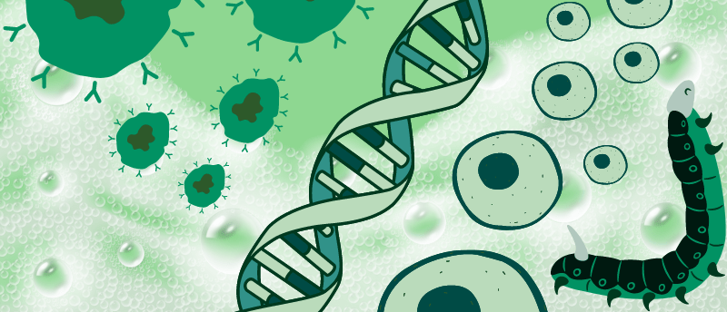
Bioengineering, the application of engineering design and analysis principles to biological systems, is an extensive field. With applications in immunotherapy, gene therapy and organ-on-a-chip models, bioengineering can be utilized in numerous areas of biomedical science, furthering our understanding of a biological system or assisting the development of a drug.
Here, we outline the recent bioengineering feats that have contributed to our understanding of the human body as well as the importance of safe data-centric research.
Nanowires preserve T cell’s naivety in immunotherapy
T-cell therapy involves the extraction of patients’ own T cells for modification in a lab before infusing them back into the patient. Although T-cell therapy has revolutionized medicine, it has limitations. During their modification, these T cells are stimulated and preactivated, which causes them to lose their naïve state. Naïve T cells are better disease fighters because they are young and more easily manipulated for certain therapeutic functions.
To overcome this limitation, researchers at Georgia Tech (GA, USA), led by Ankur Singh, have developed a method for delivering therapeutic miRNA to T cells, which pre-programs the cells while allowing them to retain their naïve state. To deliver the miRNA – a molecule that can upregulate or downregulate gene activity – the team used silicon wafer nanowires to form a needle bed. When placed on the bed, the cells were easily penetrated by the nanowires, delivering miRNA without utilizing cellular receptors and thus preserving the cell’s naïve state.
 Novel nanotube stamp system inserts proteins inside cells
Researchers have developed a stamp coated in nanoscale tubes capable of delivering proteins directly inside cultured cells with minimal damage.
Innovative materials for delivering gene therapies
Researchers at Fred Hutch Cancer Center (WA, USA) have investigated the potential of foam as a delivery method for expensive gene therapies. “Our gene therapy foam shows for the first time that by taking a small amount of an expensive gene therapy drug, increasing its volume by embedding it in a solution that is mostly made of densely packed air bubbles and then applying it to cells, we can achieve a strong and safe transfer of gene therapy agents to cells,” commented Matthias Stephan, senior author of the study. Stephan believes that the foam could be a possible solution for treating cancers or autoimmune diseases confined to certain spaces, such as ovarian or pancreatic cancer and autoimmune diseases affecting the digestive system.
To create the foam, researchers tried mixing gene therapy vectors, gene editing materials and various foaming agents, applying the concoction to cells in the lab to see if they would take up the gene edits. Testing their foam, the team made a COVID-19 mRNA solution with a genetic alteration that caused it to glow when delivered to cells, allowing the team to track its uptake. They found that the foam boosted gene transfer in the cells, outperforming liquid formulations. When the foam was injected into the belly of a mouse, the researchers observed that in only one day, the genes were transferred to the targeted cells with minimal side effects.
For more cell and gene therapy news, check out our sister site, RegMedNet.
 Designing synthetic genetic circuits to build motile ‘biobots’
Leonardo Morsut is an Assistant Professor at the University of Southern California in the Stem Cell Biology and Regenerative Medicine Department. His research group focuses on controlling multicellular behaviors using synthetic biology approaches. We spoke with Leonardo to find out about the differences between synthetic biology and classical biology approaches and his preliminary work developing a motile ‘biobot’ based on cardiomyocyte contraction.
Utilizing nature’s silk to build better organ-on-a-chip models
Organ-on-a-chip models are revolutionizing our understanding of the human body, serving as important models for drug development and disease research. However, these models are currently grown on nondegradable polymer structures, which creates a permanent barrier between the growing cells and tissues that doesn’t mimic human organs’ extracellular membranes.
To overcome this limitation, a team at Duke University (GA, USA) have developed a silk-based, ultrathin membrane that could facilitate better tissue growth. To construct their membrane, the team utilized silk fibroin, a silkworm-produced protein that can be spun into a porous membrane electronically. They tested their silk membrane by applying it to a kidney chip model, observing that the human induced pluripotent stem cells loaded into the chip could communicate through the porous silk membrane. This communication facilitated cell differentiation, producing different types of kidney cells that eventually assembled to form a glomerular capillary wall that could filter molecules by size.
 Icy veins: a new 3D printing method for building artificial blood vessels
Researchers are using ice as a template for the 3D printing of artificial blood vessels in engineered tissues, which could one day be utilized for artificial organ transplants and drug testing.
Synthetic cells follow chemical directions to change shape
Researchers at Johns Hopkins University (MD, USA) have investigated how cells initiate movement and respond to drugs by developing a synthetic cell that responds to external chemical cues and displays ‘symmetry breaking’. Symmetry breaking occurs when a cell’s molecules shift into an asymmetrical pattern just before cell movement. To mimic and control symmetry breaking, the team created a giant vesicle with a double-layered membrane – resembling a simplified cell – that was engineered to have a chemical-sensing ability, allowing the cell to respond to chemical cues and change shape. Chemical sensing could one day be used for targeted drug delivery within the body.
“The idea is that you can package anything you want into these bubbles — protein, RNA, DNA, dyes or small molecules — tell the cell where to go using chemical sensing, and then have the cell burst near its intended target so that a drug can be released,” concluded senior author Takanari Inoue.
 Could drinking carbon monoxide foam suppress tumor cell growth?
Scientists have developed a drinkable, carbon monoxide-infused foam that impacts autophagy in cancerous cells, leading to reduced cellular tumor volumes.
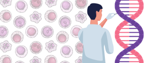
The hazards of data-centric methods in synthetic biology research
AI and synthetic biology research are becoming more and more intertwined, revolutionizing bioengineering and leading to the development of novel compounds with far-reaching applications. However, with this collaboration comes the need to think critically about the data being used to train the latest systems biology models and the access of such data for potential nefarious activity. The current study, led by researchers at the University of Bristol (UK), was built upon the work of the Data Hazards Project, which aims to outline and clarify the hazards that exist in data science research. Extending the Data Hazards Project’s work, the team set out to expand the vocabulary used to describe the safety of data-centric methods in the hopes of making future research safer.
“Having a clear vocabulary of hazards makes it easier for researchers to think proactively about what the risks of their work are and to help put mitigating actions in place. It also makes communication easier for people working across fields who sometimes use different language to talk about the same issues,” concluded co-author Nina Di Cara.
For more bioengineering news, check out our topic page.