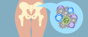
Recreating the tumor microenvironment with 3D bioprinting technology

Cancer models retaining original tumor characteristics can be developed through 3D bioprinting, allowing for rapid evaluation of drug responses.
A team of researchers led by Jinah Jang from Pohang University of Science and Technology (South Korea) and Charles Lee from The Jackson Laboratory for Genomic Medicine (CT, USA) have used 3D bioprinting technology to develop a gastric cancer model that retains a tumor’s original characteristics. This model can be used to predict individual patient drug responses within 14 days of tissue extraction.
Forecasting therapeutic outcomes is an important aspect of cancer management, with the aim of enhancing the effectiveness of anti-cancer drugs. Currently, the primary method for determining the efficacy of anti-cancer drugs is gene panel-based tests. However, even when optimal anti-cancer drugs are chosen, variations between patient responses are still seen due to tumor heterogeneity.
Although patient-derived xenografts (PDXs) and patient-derived organoids (PDOs) have significantly enhanced our understanding of cancer biology and allowed for more tailored approaches for assessing treatment efficacy, they still have some disadvantages.
PDXs require long construction times, delaying the availability of screening results. Moreover, during the passage of patient-derived cancer tissue into mice, PDXs often lose crucial stromal characteristics, resulting in alterations in the properties of the tumor microenvironment.
Similarly, PDOs – while reflective of a patient’s genomic characteristics – face uncertainties in preserving the phenotype and characteristics of the original cancer. This is due to PDOs primarily being cultured in a mouse sarcoma-derived matrix without human stromal cells.
To address these limitations, the research team utilized extrusion-based 3D bioprinting technology and bioinks laden with patient-derived tissue fragments to create an in vitro gastric cancer model that retains the original characteristics of the tumor.
 Why do some AML patients respond better to immunotherapy?
Immune cells in the bone marrow microenvironment of AML patients are key in distinguishing a response to immunotherapy.
The researchers reproduced cancer cell–matrix interactions in their model by encapsulating cancer tissues within a stomach-derived decellularized extracellular matrix hydrogel. Furthermore, cancer cell–stromal crosstalk was reproduced in the model by co-culturing the tissue with human gastric fibroblasts, which allowed the in vivo tumor microenvironment to be recreated in vitro.
Recreation of the original tumor characteristics in the model allowed for the patient response to anti-cancer drugs to be assessed with high specificity within 14 days of tissue extraction.
“By reproducing cancer cell–stroma and cell–matrix interactions, this model enhances the accuracy of drug response predictions and reduces unnecessary drug administration to non-responsive patients,” commented Lee.
The gene profile of the model closely resembled that of patient tissues for cancer development, progression and drug response, surpassing the performance of PDX models. As a result, this model could be a promising preclinical platform for predicting individual patient responses and validating new anti-cancer drugs.