Making the move from 2D to 3D cell culture: an interview with Molecular Devices’ Jayne Hesley and Jeff McMillan

Editor-in-Chief, Francesca Lake, speaks to Jayne Hesley and Jeff McMillan of Molecular Devices to discuss 3D cultures, why they are not yet in widespread use and their future.

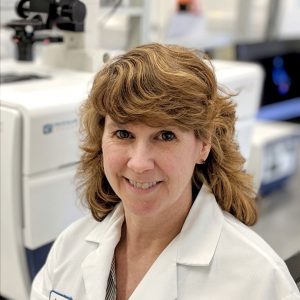
Jayne Hesley
Jayne Hesley is a Senior Applications Scientist for Cellular Imaging at Molecular Devices, LLC. She has over 10 years’ experience developing cell-based applications using ImageXpress Micro high content imaging systems and MetaXpress automated analysis software. Most recently her focus has been on exploring the use of 3D models, particularly cells or spheroids cultured in hydrogels, for studying cell viability, apoptosis, morphological changes, mitochondrial damage and autophagy. These studies include the use of automated confocal microscopy for live-cell timelapse experiments as well as screening assays in microplates. Prior to working in the imaging field, Jayne developed colorimetric and fluorescence assays, including TR-FRET and fluorescence polarization, for high-throughput screening on multi-mode readers. She has a B.S. in Animal Science and an M.S. in Biology.

Jeff McMillan
Jeff McMillan is a Senior Product Manager supporting the continued development of Molecular Devices’ new imaging product lines with a specialization in software for the ImageXpress platforms. Jeff brings over 20 years of experience managing life sciences instrumentation and software products for companies ranging from Agilent to Thermo Fisher. Jeff’s education in physics from UC Berkeley and MBA from Santa Clara has given him a solid foundation to launch many hardware and software solutions into the marketplace.
The advantages of 3D cell culture and imaging are fairly well understood, particularly in drug discovery and development, but 2D culture and imaging are still common in labs – why is this?
Jayne Hesley: Scientists know that 3D better represents the biology of the organism they are trying to study but, at this point, 2D imaging experiments are well-characterized, relatively simple, and often will answer the question the scientist is asking. We’re just at the tipping point for more widespread 3D use in labs; the default today is 2D.
Jeff McMillan: 2D experiments are how we all learned in school, and so it stands to reason that nearly all laboratories continue to perform 2D experiments. I’d imagine as much as 40% of laboratories have some 3D imaging equipment, but that number is growing very fast. We hear from new and existing customers that they are incorporating or even expanding 3D imaging capabilities in their labs because it enables faster discovery and greater understanding. We’re absolutely seeing strong growth in that area.
What options are there to help researchers overcome the complex challenges that come with this field and perform accurate and reproducible 3D cell culture research?
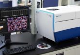
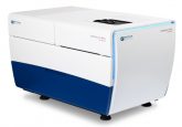
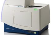
Jayne Hesley: Practically speaking, flexibility, innovative tools and true 3D analysis are three ways researchers can perform accurate and reproducible 3D cell culture research. One of the biggest challenges is that 3D samples are available in a variety of labware and require specialized slides. There is currently little standardization. This is where the instruments used in a laboratory are so important; Molecular Devices’ imaging instruments have the flexibility to acquire images in non-standard formats, sizes and spheroids.
Second, as 3D cell culture use increases, so do challenges in both acquisition and analysis – particularly as the physical size of the 3D objects increase. Scientists need tools that can handle this challenge. Innovations like water immersion objectives can both minimize the optical distortion of the object being imaged as well as permit the collection of more light, thus allowing us to peer deeper into the sample.
Third, our software has true 3D analysis: with it, you can create a 3D image by connecting various data points, identifying markers and measuring objects that may even span over multiple planes – all to give comprehensive information about the cells or spheroids. These analyses can be paired with live cells over time. Using 3D analysis of the entire volume, scientists have been able to trace and measure the penetration of T cells into a 3D tumor model, helping identify compounds that might weaken the tumor in ways not possible with 2D. That’s very exciting!
What challenges still remain in this field?
Jeff McMillan: Helping researchers overcome the move from 2D to 3D is at the forefront of our minds at Molecular Devices, and it shapes our internal R&D as well as our collaborations with companies such as Mimetas and Stemonix. We’re scientists too, and we are constantly thinking through the entire laboratory ecosystem and experiment lifecycle to lower hurdles, close gaps and align our products with other instruments that would already be in a lab. 3D imaging may be complex, but deploying these instruments in a lab doesn’t need to be.
Ultimately, I believe ease of use, flexibility and openness to collaborate with other companies is quickly lowering the barriers to widespread use. I’m proud that Molecular Devices is leading an effort to make moving to 3D easier for researchers.
What are your respective visions for the future of this field?
Jayne Hesley: Drug screening, especially using tumor models, will be within reach of many labs with standardized spheroid generation and assays. The continuing development of more complex models such as organoids will lead to large studies that will show their predictive value and could substantially reduce live animal testing. Personalized medicine is another new frontier accelerated by 3D culture, and will change how we administer patient care – and cures.
Jeff McMillan: I see an upcoming revolution in data analysis. With the advent of tools like machine learning and artificial intelligence already being explored by both users and solution providers, the wealth of data being generated by imaging systems will be more effectively analyzed and will lead to studies with higher predictive powers.
Read more on Molecular Devices here.


