3D-printed lungs provide an insight into respiratory diseases

In a recent study, researchers developed artificial lungs that will enhance the efficiency of respiratory disease research.
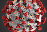
A collaboration between researchers at Pohang University of Science and Technology (POSTECH; South Korea) and the Korea Research Institute of Chemical Technology (KRICT; Daejeon, South Korea) has built artificial lungs to study infections and test possible drugs for respiratory diseases, including COVID-19. Their 3D-printed lung was designed in the hopes of reducing the time and money needed to progress drug development for respiratory diseases.
COVID-19 spurred scientists all over the world into action, demonstrating mass mobilization against the virus and the search for an effective treatment. The researchers at POSTECH and KRICT were not immune to this mobilization and set about developing artificial lungs to make the study of respiratory diseases, and the development of their potential treatments, more efficient.
The research team used 3D printing technology to develop an artificial lung composed of a vascular epithelium, extracellular matrix and epithelium, mimicking the respiratory tract tissue structure of humans. The 3D model also displayed functional capabilities of the human respiratory tract, with working cell–cell junctions and mucus secretion. They developed their model to include proteins in the epithelial layer that are known entry points for COVID-19, namely ACE2 and TMPRSS2. Unlike previous 2D cell models infected with the virus, this model lasted 21 days post-infection, allowing the researchers to monitor and understand the virus’ progression and effects on the lungs.
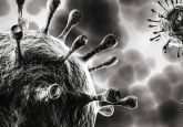
How Pseudomonas aeruginosa penetrates the lungs
Scientists now know how Pseudomonas aeruginosa infects the lungs thanks to human stem cell-derived lung microtissues.
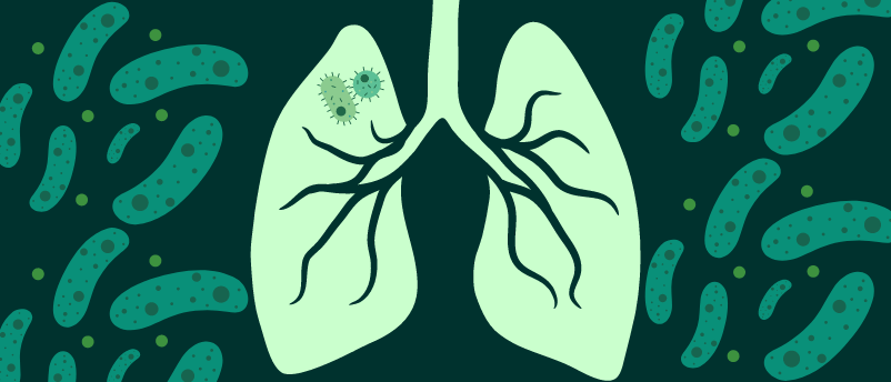
During these 21 days, the researchers observed infection-induced cell lesions and barrier degradation; using in-depth transcriptomic analysis, the researchers were able to identify dramatic gene expression changes affecting the infection pathway, host immune response and viral proliferation. All these observations were consistent with data collected from COVID-19 patients.
What’s more, the team was able to perfectly model how two COVID-19 drugs – remdesivir and molnupiravir – reach the infection sites on the epithelial layer to inhibit viral replication. Their model demonstrated how these drugs travel across the tissue barrier to reach the epithelial layer, a process not observed in 2D models where drugs are applied directly to the epithelial cells. This allowed them to verify treatment efficacy, calculate appropriate dosages and determine potential side effects.
Their 3D model has shown significant promise as a human-like model for lung function and respiratory drug testing, offering a more efficient and effective disease-modeling and treatment-testing research tool.