Immunohistochemistry and immunofluorescence: adapting to the needs of life science researchers

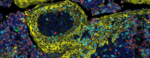
Immunohistochemistry is a protein-detection technique that uses antibodies to identify antigen markers present in a tissue sample. The antibodies are tagged with enzymatic labels, which activate when an antibody binds to an antigen. This allows the antigens to be visualized under a microscope. Immunofluorescence works much like immunohistochemistry, except instead of an enzymatic tag, it uses a fluorescent signal. Both techniques are widely used; here, we highlight recent applications of and adaptations to these techniques and their methodologies that have enabled them to meet the growing needs of life science researchers.
Immunohistochemistry (IHC)
Tumor immune characterization in breast cancer
Researchers from Lund University (Sweden) have investigated the tumor immune microenvironment of primary triple-negative breast cancer using digital image analysis of IHC-stained immune cells. To do this, they developed tissue microarray marker quantification (TMArQ) – an open-source image analysis pipeline – and applied it to IHC stains for immune markers in cancer tissue. Alongside this analysis, the team conducted whole-genome and RNA sequencing. TMArQ demonstrated that immune markers are co-expressed in the tumor immune microenvironment. The team was able to show that when TMArQ is combined with phenotypic analysis, it can improve our understanding of the molecular mechanisms underlying the tumor immune microenvironment.
How mosquito saliva facilitates malaria infection
A research team from the National Institute of Health (MD, USA) has utilized IHC and other techniques to determine the function of Anopheles gambiae salivary apyrase (AgApyrase) in regulating hemostasis – the process that stops blood from coagulating – in mosquitos’ blood meal and during transmission of Plasmodium, the parasitic protozoan that causes malaria, from a mosquito to a host. They performed IHC using anti-AgApyrase antibodies on dissected human blood-fed mosquitoes and unfed mosquitoes, which confirmed that mosquitoes ingest a significant amount of apyrase – a known inhibitor of platelet aggregation – during feeding. The team utilized IHC along with other techniques to discover that apyrase activates tissue plasminogen activator, which causes a cascade of interactions leading to Plasmodium transmission.
IHC meets deep learning
IHC images are essential for diagnosing disease and can help produce improved computer-aided diagnostic systems. In a recent study, researchers at Wenzhou Polytechnic (Zhejiang Province, China) developed a novel technique to generate high-quality IHC-stained images. This technique is called harmonic conditional generative adversarial network (HcGAN), and it relies on hematoxylin and eosin (H&E) images that depict cellular and morphological structures in cancer tissues. By combining two staining techniques, IHC and H&E, researchers not only glean information on specific protein markers that assist with tumor classification, but also obtain clear visualizations of tissue structure, cell distribution and morphological changes.
 Antibody validation for successful immunohistochemistry and multiplex immunofluorescence
This webinar describes how to evaluate and select the correct monoclonal antibody for use in mIF assays and introduces a simplified approach to optimize antibodies for use in an immune checkpoint multiplex assay, resulting in half the time typically spent on validating multiplex panels.
Immunofluorescence (IF)
Using IF to capture cell states
The regulation of cell states is dependent on signal cascades predominantly stimulated by receptor–ligand binding on the cell membrane and the interactions that occur between cells. Multiplexed IF enables the investigation of membrane proteins by visualizing signal activation and providing a spatially resolved picture of the membrane proteins. In this study, researchers from Kyushu University (Fukuoka, Japan) developed precise emission-canceling antibodies (PECAbs), which have cleavable fluorescent labels. PECAbs allow researchers to conduct high-specificity sequential imaging utilizing hundreds of antibodies, expanding the capabilities of IF to provide temporal information in addition to its spatial prowess. The researchers showed that by combining the spatiotemporal data of signaling pathways obtained by PECAb-based IF with those collected using seq-smFISH, they were able to classify cells and their states in human tissue, enabling complex cell process analysis.
Dual-modality imaging for whole-slide analysis
In this study, an international collaboration of researchers combined high-resolution IF and high-dimensional imaging mass cytometry (IMC) on the same tissue slide to develop a highly practical dual-modality imaging method. Their computational method uses an IF whole-slide image to establish a spatial reference before integrating IMC. IF enables accurate single-cell segmentation that allows researchers to extract high-dimensional IMC features for further analysis. The team applied their dual-modality imaging method to esophageal adenocarcinoma of different stages, revealing a single-cell pathology landscape of the cancer.
Automated IF pattern recognition in autoimmune diseases
AI is fast becoming a diagnostic tool, with researchers integrating machine and deep learning into image analysis and symptom reporting. However, AI has yet to reach IF imaging for autoimmune bullous skin diseases (AIBDs). Direct IF assigns fluorophore-labeled antibodies to specific target proteins fixed on patient skin tissue. Direct IF, although a useful diagnosis tool, requires manual interpretation, which can significantly hinder efficiency. To remedy this, researchers from the University of Genoa (Italy) have investigated the potential of deep learning in automating the classification of direct IF patterns. By automating this process, the researchers believe that AIBD diagnosis will benefit.