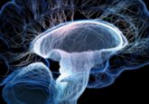
Mapping the aging process

Spatial maps of various tissues and organs in aging mice have revealed new insights into the aging process.
Aging is a process common to many animals; however, the complexity of aging means that there’s much we still don’t know about it. Now, researchers from the Chinese Academy of Sciences (Beijing, China) and BGI Research (Guangdong, China) have developed spatial maps of various tissues and organs in aging mice. The maps have provided new insights into key processes seen in aging, including ‘aging hotspots’ and the role of antibodies, and opened the door to developing therapeutics to address these processes.
Loss of tissue integrity and organ dysfunction are key characteristics of aging. However, what drives these changes is still unclear. To investigate these processes, the researchers used spatial transcriptomics to create maps of nine tissues – hippocampus, spinal cord, heart, lung, liver, small intestine, spleen, lymph node and testis – in male mice during aging, detailing the spatial distribution of over 70 cell types.
The researchers then used a novel method called organizational structure entropy analysis and found that increased spatial structural disorder and a loss of cellular identity are universal indicators of systemic aging. This suggests that spatial structural damage may be a primary cause of functional decline in organs as they age.
 Protein aggregation pathway uncovered in ALS
The pathway leading to the telltale sign of ALS, protein aggregation in the cytoplasm of motor neurons, has been revealed.
The team also used the maps to identify senescence-sensitive spots (SSSs), which are structural regions in tissues that are more vulnerable to the effects of aging. They found that areas closer to SSSs display higher levels of tissue structural entropy and greater loss of cellular identity.
In immune organs, plasma cells, which produce antibodies, and cells with specific structures and functions are the main components of the SSS microenvironment. The expression of antibody-related genes was also increased in cells around SSSs.
In further experiments, the researchers found an accumulation of immunoglobulin G (IgG), a type of antibody, in multiple tissues and organs from aged mice and human samples, suggesting the accumulation of antibodies as an aging hallmark and the potential of IgG as a biomarker of aging.
In human and mouse macrophages and microglia, IgG was found to directly induce aging by releasing inflammatory factors. Injecting IgG into young mice induced aging in multiple tissues and organs, while reducing IgG using antisense oligonucleotides in mice delayed aging across various tissues.
The study is the first to map the spatial transcriptome of multiple organs and tissues in aging mammals and opens the door to finding new strategies to address the loss of tissue integrity and organ dysfunction seen in aging animals.

