Mitochondria of memory: the mitochondrial process supporting social recognition
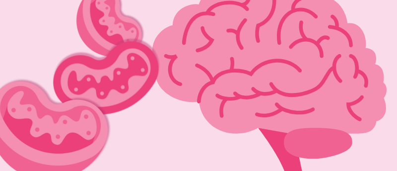
This mitochondrial process supports the brain cells essential to learning, memory and social recognition.
We know that mitochondria are the powerhouses of the cell, but what role do they play in strengthening neuronal connections? Now, researchers at the Fralin Biomedical Research Institute at Virginia Tech (VA, USA) have identified a mitochondrial process essential to brain cells involved in learning, memory and social recognition, which could have implications for Alzheimer’s, among other neurological disorders.
Although it has long been clear that mitochondria play an important role in cellular functioning, the extent to which mitochondria influence cell-specific functions remains largely unknown. Mitochondrial dysfunction has been observed and reported in neurological disorders such as Alzheimer’s, where outermost synapses are among the first synaptic connections affected, which led the current research team to investigate the connection between mitochondria and neuronal function.
They focused on the CA2 region within the hippocampus, a specialized area in the brain’s memory center that is critical for social recognition – the ability to remember and distinguish people. Interestingly, unlike surrounding hippocampal regions, the neurons in the CA2 region resist certain forms of synaptic plasticity, which is the process of strengthening neuronal connections fundamental to cognitive function and adaptive learning. This resistance raised questions about their specialized function.
Previous research has indicated the mitochondrial calcium uniporter’s (MCU) role in coupling neuron activity and mitochondrial ATP production; the MCU is a protein that regulates calcium flow into mitochondria. It has been observed that MCU is enriched in CA2 distal dendrites compared to proximal dendrites. It has also been shown that synapses onto CA2 distal dendrites readily express plasticity, whereas the synapses onto CA2 proximal dendrites resist plasticity. However, the mechanisms underlying these differences in plasticity are unknown.

New method to study gut alterations in Alzheimer’s disease
X-ray tomography can be applied to identify cellular and structural alterations of the gut, which could be pivotal for early detection of Alzheimer’s disease.
To explore this, the team used an MCU-deletion mouse model and evaluated synaptic plasticity at different locations along neurons. They found that this disrupted the plasticity of synapses further from the soma while leaving those connections closer to the soma unaffected. “This suggests that mitochondrial diversity isn’t just a biological quirk,” commented lead author Shannon Farris. “It’s a fundamental feature that allows different parts of the same neuron to function in distinct ways.”
Taking their research a step further, the team used scanning electron microscopy and AI to identify how MCU depletion affects dendritic mitochondrial morphology and content. They mapped the mitochondrial structure in CA2 neuron dendrites of both control and MCU-depleted mice at high spatial resolution with extreme precision over millimeter expanses of tissue. The analysis revealed that MCU-deficient mitochondria were smaller and more fragmented, a structural shift that may underly their impaired ability to support synaptic function.
“These findings challenge the long-held assumption that mitochondria function uniformly within dendrites,” highlighted Katy Pannoni, first author of the study. “Instead, our work suggests that mitochondria are highly specialized to support the distinct needs of different neural circuits.”
The research contributes to our understanding of mitochondrial dysfunction and its link to synaptic plasticity and therefore neurological disorders, opening new pathways to consider for potential therapies. In the future, Farris and her team plan to explore how mitochondria in CA2 neurons develop their specialized properties and delve deeper into therapeutic strategies that could support mitochondrial health, and in turn, brain health.