Protective brain cells successfully transplanted in mice without immune-suppressing drugs

New hope for the utilization of brain cell transplantation for the repair of damaged brain tissues.
Researchers at Johns Hopkins University School of Medicine (MD, USA) have demonstrated a new approach for transplanting brain cells. Their method exploits the immune system of the transplant receiver to ensure the cells are not rejected, without the need for immune-suppressing drugs.
The body’s response to foreign tissue is a major hurdle to overcome when transplanting cells, tissues or organs. Immune-suppressing drugs dampen the immune response as a whole leaving patients open to the risk of infection. Another drawback of these drugs is that patients will need to take them indefinitely.
To overcome the immune response the team sought to manipulate T cells in order to stop them from recognizing the foreign cells and initiating an immune response. They utilized CTLA4-Ig and anti-CD154, two antibodies that regulate T cells.
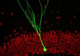
The team transplanted glial-restricted progenitor (GRP) cells into mouse brains that had been genetically engineered to lack glial cells. GRPs arise from neuronal stem cells and are a protective brain cell that can migrate and myelinate previously dysmyelinated cells. The GRPs were tagged with green fluorescent protein in order to image the transplanted cells.
After transplantation the mice received the antibody treatment for 6 days. In control group mice who did not receive the antibody treatment, there was no imageable trace from the green fluorescent protein after 21 days, indicating that the GRPs did not survive. In mice who received the antibody treatment there was a detectable glow for more than 203 days, demonstrating that even after treatment ceased the GRPs were not destroyed by immune cells. Next, the team assessed whether the GRPs were able to carry out their normal function and restore myelination. Utilizing MRI, they assessed structural differences between mouse brains with normal glial function and those without. It was demonstrated that myelin was populated in the brains of mice who had received the GRP transplant and the antibody treatment, confirming that the transplanted cells were able to commence neuronal protection.
- Source of new neurons for the hippocampus
- New therapy for neurodegenerative disorders
- Novel drug combination converts glia to restore neuron function
This study gives hope for the treatment of children who suffer from genetic diseases in which myelin does not form properly. One such disorder is Pelizaeus–Merzbacher disease, which affects approximately one in every 100,000 children born in the USA. The condition is life altering and affects children’s motor skills, resulting in them missing developmental milestones. It also causes involuntary muscle spasms and sometimes partial paralysis of the extremities.
“Because these conditions are initiated by a mutation causing dysfunction in one type of cell, they present a good target for cell therapies, which involve transplanting healthy cells or cells engineered to not have a condition to take over for the diseased, damaged or missing cells,” commented study lead Piotr Walczak.
These findings are preliminary, with focus solely on localized regions of the brain. In the future the team aims to combine their discovery with cell delivery methods to ensure global repair of brain tissues.


