Regulatory mechanism of centromere distribution uncovered

Researchers propose a two-step regulatory mechanism for non-Rabl configurations of centromeres in the nucleus.
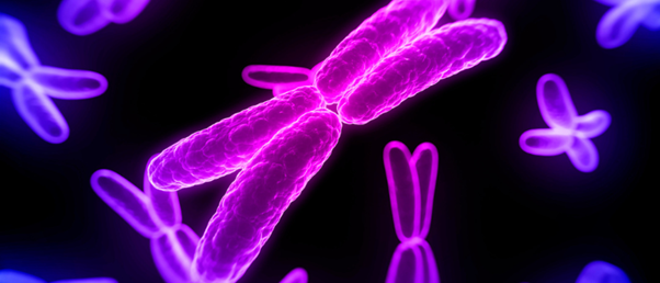
Centromeres are chromosomal domains that link pairs of sister chromatids together during cell division. When cells divide, centromeres are pulled to opposite ends of the cell. Once divided, centromeres are distributed within the nucleus in either a Rabl or non-Rabl configuration, named after the 19th century cytologist, Carl Rabl. In the Rabl configuration, the distribution of centromeres remains unchanged as they are grouped to one side of the nucleus, while in the non-Rabl configuration, centromeres are dispersed throughout the nucleus.
The biological function and molecular mechanism of Rabl and non-Rabl configurations have remained a mystery since the 1800s. Now, a collaboration led by researchers at the University of Tokyo (Japan), along with other institutions in Japan and Switzerland, has uncovered the molecular mechanism behind non-Rabl configurations.
Using cytogenic and molecular analysis, the researchers studied a plant known to have a non-Rabl configuration of centromeres called Arabidopsis thaliana, also known as thale cress, as well as a mutant of this plant with a Rabl configuration. They found that two protein complexes work together to determine the centromere distribution during cell division: condesin II (CII) and the linker of nucleoskeleton and cotoskeleton (LINC).
“The centromere distribution for non-Rabl configuration is regulated independently by the CII-LINC complex and a nuclear lamina protein known as CROWDED NUCLEI (CRWN),” explained Sachihiro Matsunaga (University of Tokyo), the senior author of the paper.
The researchers proposed a two-step mechanism for non-Rabl distribution of centromeres. First, the CII-LINC protein complex mediates centromere scattering from late anaphase to telophase (two phases towards the end of the cell division mechanism). This is then followed by the second step; CRWN stabilizing the scattered centromeres on the nuclear lamina, which is a protein mesh attached to the inner nuclear membrane, within the nucleus.
 Streamlining image analysis for mitotic cells with AI
Researchers developed a deep-learning model for artificial intelligence (AI) to recognize mitotic cells, which is easy-to-use, easy-to-train and aimed at non-data scientists.
Next, the researchers investigated the biological significance of centromere distribution and analyzed the gene expression in Arabidopsis thaliana and in the Rabl-structure mutant. The researchers hypothesized that the spatial arrangement of centromeres also changes the spatial arrangement of the genes, so hoped to observe changes in gene expression between the Rabl and non-Rabl plants. They did not observe this. However, they did find that when DNA damage stress was applied, the Rabl-mutant plant grew organs at a slower rate than the unaltered plant.
“This suggests that precise control of centromere spatial arrangement is required for organ growth in response to DNA damage stress, and there is no difference in tolerance to DNA damage stress between organisms with the non-Rabl and Rabl,” said Matsunaga. “This suggests that the appropriate spatial arrangement of DNA in the nucleus regardless of Rabl configuration is important for stress response.”
Matsunaga revealed that the next steps will be to identify the power source that changes the spatial arrangement of specific DNA regions and the mechanisms that underlie this process.
“Such findings will lead to the development of technology for artificially arranging DNA in the nucleus in an appropriate spatial arrangement,” explained Matsunaga. “It is expected that this technology will make it possible to create stress-resistant organisms, as well as to impact new properties and functions by altering the spatial arrangement of DNA rather than editing its nucleotide sequence.”