Photoacoustic tomography breakthrough could transform vascular imaging diagnostics

Researchers have developed a new photoacoustic tomography scanner that can produce high-quality images of vasculature and microvasculature in seconds.
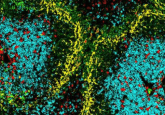
Photoacoustic tomography (PAT) has been around since the 2000s; however, the time it takes to produce images has prevented its widespread clinical use. Now, a group of researchers from University College London (UK) have developed a new PAT scanner that can acquire images of vasculature and microvasculature 100 to 1000 times faster than previous scanners. This makes PAT a suitable technique for diagnosing and monitoring conditions that affect vasculature, such as cancer and diabetes.
For some conditions, like diabetes and inflammatory skin diseases, sub-cm visualization of microvasculature is required for clinical assessment. PAT presents one solution for this visualization and involves directing short laser bursts at a tissue, some of which are absorbed, leading to a slight increase in heat and pressure that generates a faint ultrasound wave containing information about the tissue. By capturing these waves at the skin surface, an image of the vasculature can be created.
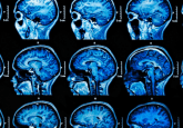
Although PAT can successfully visualize veins and arteries up to 15mm deep in human tissues, historically the technique has been too slow to produce high-enough quality 3D images for use by clinicians. Patients undergoing a PAT scan must remain completely still, as any movement can blur the images. Expecting patients to stay motionless for over 5 minutes is impractical, and the resulting images may not always be clinically useful. Reducing the time it takes for PAT scanners to capture an image would minimize motion-induced blurring, enhance image quality and make the technique more practical.
Earlier PAT scanners measured ultrasound waves at more than 10,000 different points over the tissue surface, one at a time. To reduce the time needed to acquire images, the researchers developed a PAT scanner that detects ultrasound waves at multiple points simultaneously. They also used similar mathematical principles to those used in digital image compression to reconstruct images from a few thousand measurements of the ultrasound wave, rather than tens of thousands. Combined, these developments enabled the scanner to take images in a few seconds or even milliseconds, delivering high-quality 3D scans to clinicians in real time and allowing the visualization of dynamic changes in the tissue.
 AI for an eye: AI speeds up retinal imaging
Researchers have developed an AI tool that makes retinal imaging 100 times faster.
The team then tested their scanner on ten patients with type-2 diabetes, rheumatoid arthritis or breast cancer, as well as seven healthy volunteers. They found the scanner could provide detailed 3D images of vasculature at various locations and show disease-associated vascular changes, features that can’t be seen using conventional imaging techniques like MRI.
For example, in three patients with type-2 diabetes, the scanner showed changes in the microvasculature in the feet, an indicator of peripheral vascular disease (low blood flow to the feet and legs), a complication often experienced by people with diabetes.
“In one of our patients, we could see smooth, uniform vessels in the left foot and deformed, squiggly vessels in the same region of the right foot, indicative of problems that may lead to tissue damage in future,” commented co-senior author Andrew Plumb. “Photoacoustic imaging could give us much more detailed information to facilitate early diagnosis, as well as better understand disease progression more generally.”
Cancer also displays changes in microvasculature, with tumors having a high density of small blood vessels compared to other tissue. PAT could therefore be used to detect and monitor tumors and to distinguish between tumor tissue and normal tissue during and after surgery.
The developments make PAT suitable for clinical use for the first time; however, the researchers stress that the scanner needs to be tested on larger groups of patients to confirm their findings before moving to the clinic.