Molecular Devices unveils next-generation imaging system with deep learning technology

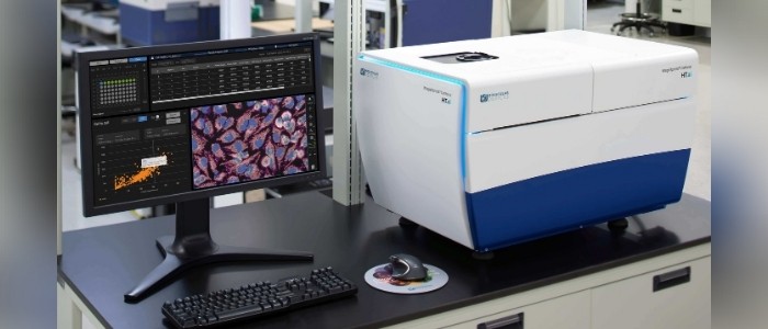
San Jose, CA, USA., Jan. 25, 2021 – Molecular Devices, LLC., a leading provider of high-performance life science technology, has globally launched its next-generation, high-content imaging system. Built on the success of the company’s flagship model, the ImageXpress® Confocal HT.ai High-Content Imaging System is designed to help researchers advance phenotypic screening of 3D organoid models. The new system offers faster image acquisition and multiplexing flexibility with high-performance lasers. Image analysis with machine learning will enable researchers to quickly uncover new insights that are provable and repeatable with clear, accurate data.
“As a trusted partner to scientists for over 35 years, Molecular Devices relentlessly innovates high-content imaging and analysis technology that simplifies complex biological research leading to faster, more reliable scientific discoveries,” said Susan Murphy, President of Molecular Devices. “The ImageXpress Confocal HT.ai system with self-learning image analysis software follows a long history of life science solutions that empower our customers to solve their most challenging research questions with the utmost confidence.”
Solving 3D biology challenges with high-throughput imaging and analysis technology
The ImageXpress Confocal HT.ai system offers up to double the throughput speed for 3D organoids and spheroids while helping the scientific community overcome roadblocks when working with complex cell models. Notably:
- Increased multiplexing flexibility due to high-performance laser excitation with seven laser lines and eight filter combinations.
- More accurate and reproducible image analysis enabled by new dual micro lens spinning disk confocal technology with enhanced field uniformity.
- Greater image resolution and sensitivity with up to 4X increase in signal leading to lower exposure times with automated water immersion objective technology.
- Improved accuracy and robustness of high-content image analysis with machine learning through IN Carta™ Image Analysis Software. Integrated from Cytiva – formerly part of GE Healthcare Life Sciences and now a Danaher company – the software has an intuitive guided workflow in a modern user interface, and uncovers data insights that other technologies miss.
Reducing the burden on researchers with machine learning
Turning high-content images into actionable insights is a complex process. Relying on traditional analysis methods can lead to errors or oversimplifications that can skew results and diminish assay reliability. IN Carta software paired with the ImageXpress Confocal HT.ai system uses machine learning to easily convert data-rich image information into repeatable results, while reducing the burden on researchers.
Two key components of IN Carta software that leverage machine learning are the software modules SINAP and Phenoglyphs. SINAP uses deep learning to detect objects of interest to improve accuracy and reliability in the first step of image analysis. Phenoglyphs takes hundreds of image descriptors extracted by SINAP and creates an optimal set of rules for grouping objects with a similar visual appearance. Both modules utilize unsupervised decisions to generate an initial result that is iteratively optimized though user input. Together, they improve the integrity and accuracy of findings through an easy-to-use, end-to-end workflow.
Showcasing innovation at SLAS 2021
Molecular Devices will feature the ImageXpress Confocal HT.ai system with IN Carta software in the “Product Showcase” section of the company’s virtual booth during the SLAS 2021 online tradeshow. Through video chat, attendees will also have the opportunity to interact one-on-one with product experts and applications scientists, discussing ways to maximize research efforts with the company’s advanced life science solutions.
Additional supporting resources will include the video presentation, “Capturing complexity of 3D biology: New advantages of high-content imaging,” scheduled for Wednesday, January 27, at 5 p.m. EST, as well as the following ePosters:
- Using laser light source for high-content automated imaging to improve sensitivity, speed, and precision for complex biological assays
- High-content imaging of 3D lung organoids as an assay model for in vitro assessment of morphology and toxicity effects
- Simplified workflow for phenotypic profiling based on the cell painting assay
This content was supplied by Molecular Devices.


