Advancements and applications of tumor organoids in cancer research
Organoid models of patient-specific tumors are transforming our understanding of cancer heterogeneity and its implications for personalized medicine. In this interview, Janina Moros, Alexander Trampe and Zinnia Noor, who all work for PromoCell (Heidelberg, Germany), discuss the benefits of tumor organoids, how to establish and culture them and their applications across different areas of cancer biology.
 Janina Moros studied at the University Tübingen (Germany). She did a bachelor’s degree in biochemistry and master’s in molecular cell biology and immunology, focusing on human genetics and neurodegenerative disorders. Janina’s master’s thesis at the German Cancer Research Center (Heidelberg, Germany) was on the secretome of breast cancer cell lines and the influence on human primary endothelial cells. Janina started at PromoCell in July 2022 as a Scientific Support Specialist, and helps scientists navigate PromoCell’s product portfolio, troubleshoot challenges and optimize their cell culture workflows.
 Zinnia Noor studied biochemistry and molecular biology at the University of Dhaka (Bangladesh). She obtained a master’s degree in biotechnology from the University of Kaiserslautern (Germany), now known as the University of Kaiserslautern-Landau. Later, Zinnia obtained her PhD from the same university in a collaborative project with Saarland University (Germany) and University of Alberta (Canada). Her project involved the investigation of the contribution of an electrogenic membrane transporter in the brain in sodium homeostasis along with Na-K-ATPase under different physiological conditions. Zinnia works at PromoCell as Scientific Support Specialist and uses her cell culture experience to support customers in their research endeavor and provide product and service-related consultation.
 Alexander Trampe studied biochemistry at the University of Bielefeld (Germany) and completed his PhD in cell biology at the University of Lübeck (Germany). After over 10 years of experience as a Scientific Support Specialist, Alexander is now the Project Manager of PromoCell’s Cancer Media Toolbox.
In cancer research, which features of tumor organoids are advantageous over traditional 2D cell culture?
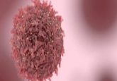
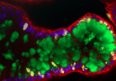
Tumor organoids offer a more physiologically relevant model of cancer than 2D cultures. This is because tumor organoids are 3D structures that closely resemble tumors in organization and cell types. As a result, they allow for realistic cell-to-cell interactions and signaling pathways. Thereby, organoids serve as a powerful bridge between conventional in vitro models and in vivo physiology of the human body, while 2D cultures are too simplistic to mimic the complex environment of a tumor.
Derived from primary isolates of patient samples, tumor organoids provide a personalized model, preserving the genetic and functional diversity of a patient’s tumor. Organoids capture tumor heterogeneity, reflecting a mix of different cell types, which enables the study of interactions and contributions to cancer progression. Organoids offer greater accuracy in drug testing compared to 2D cultures, bridging the gap between promising results and actual patient response, making them a more reliable platform for testing new drugs.
In your opinion, what are the best methods currently in use to establish tumor organoids from patient-derived samples?
When generating organoids from patient samples, the aim is to closely replicate tumor characteristics in vivo. The dynamic interaction between cancer stem cells (CSCs) and the tumor environment supports CSC growth and maintenance, crucial for tumor development and progression, including metastasis. Therefore, it’s essential to select a system for establishing tumor organoids from patient-derived samples that supports CSCs, allows co-culture of cancer and non-cancer cells, and preserves the tumor microenvironment. A patient’s tumor is unique and tumors from different patients have different characteristics. Accordingly, it is important to choose a system that preserves the malignancy as well as the individual’s genetic and functional characteristics of the tumor.
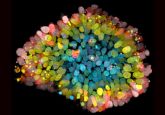
While the use of an extracellular matrix (ECM) is well established, generating 3D cancer models without one, such as with the 3D Tumorsphere Medium XF, can enforce CSC traits by selecting for anoikis-resistance and additional stem-cell properties. Tumorspheres form through the proliferation of a single CSC, with undifferentiated CSCs in the core and more differentiated cells at the outer borders. Furthermore, co-culture, such as with fibroblasts, is feasible in this setting, as they can integrate into the tumorspheres predominantly composed of cancer cells.
This approach allows for the establishment of a more differentiated, in vivo-like ‘tumor-organoid-type’ setting, while also allowing a CSC-focused tumorsphere culture. Thus providing a customizable approach to address various research needs.
Once established, what are the specific media and growth conditions required for optimal tumor organoid maintenance?
When establishing organoid cultures, it is crucial to choose a medium that supports CSCs, differentiated cancer cells and non-cancerous cells to preserve tumor viability and functionality. Preserving the viability and functionality of CSCs and therefore the malignancy of the tumor, allows researchers to focus on this critical cell population and its role in tumor biology without bias from cancer cell-specific markers.
Maintaining long-term viability of organoids is challenging. However, using media that supports tumor-initiating cells like CSCs can significantly enhance viability. Optimal conditions include a 3D medium that supports CSCs while maintaining tumorigenicity. This allows for the cultivation of various cell types, facilitates complex spheroid formation, and enables reproducible homogeneous tumorsphere formation, scalability for >10 passages and a defined synthetic growth medium composition.
What challenges are associated with culturing tumor organoids and how can researchers overcome them to ensure consistent and reliable results?
As discussed earlier, tumors show significant heterogeneity between patients and within a single tumor. Further, to generate a reliable 3D tumor model, the complex network of signals and interactions between tumor cells and the tumor microenvironment (e.g. blood vessels, immune cells) needs to be replicated in your organoid culture, making it a highly complex cell model. To address these challenges, using a medium that captures this diversity while allowing a standardized culture system is crucial. Optimized media supporting the metabolism of malignant cells like CSCs can enhance long-term organoid viability.
Experience has shown that the success of obtaining suitable 3D cancer models from biological material can be inconsistent, often due to the absence of CSCs or low tumor malignancy in the sample, rendering it unsuitable for proper assays and suggesting potential sample discard.
Moreover, establishing and culturing organoids is often labor-intensive and requires specialized products, limiting the scalability for high-throughput drug screening or large-scale studies. Automating steps and developing new techniques for generating large numbers of organoids need to be explored to improve scalability.
How can tumor organoids be used to study different aspects of cancer biology?
Tumor organoids offer immense promise for drug discovery and development by accurately representing tumor physiology and retaining genetic characteristics. Organoid-based drug testing can be used for a more accurate prediction of how a drug might work in vivo, which can help identify promising drugs early on and avoid wasting time and resources on drugs that may not be effective in vivo. Personalized approaches to drug screening can lead to the development of more targeted and effective treatment plans that are tailored to the patient’s tumor. By assessing how a patient’s tumor cells respond to a panel of drugs, doctors can choose the therapy with the highest prediction efficiency, potentially leading to improved treatment outcomes and reduced side effects. Furthermore, by exposing organoids to drugs over time and observing how they adapt, researchers can gain insights into resistance mechanisms and develop strategies to overcome them and thus improve the effectiveness of existing therapies.
The tumor microenvironment is a critical player in cancer progression. It consists of various cell types like immune cells, endothelial cells, which form blood vessels, and stromal cells, which provide structural support. These cells interact with cancer cells through a complex network of signaling molecules and influence tumor growth, invasion and metastasis. The interplay between cancer cells and the tumor microenvironment is crucial for understanding how the microenvironment influences cancer. Organoid models that integrate the tumor microenvironment provide a more realistic model that can be used to understand cancer biology and drug resistance. Metastasis is a major hurdle in cancer treatment. Organoid models can be used to study the process of metastasis by observing how tumor cells detach, migrate and invade other tissues. This deeper understanding can lead to the development of therapies that target specific steps in a metastatic cascade and prevent cancer from spreading.
Overall, tumor organoids represent a powerful emerging technology with the potential to transform cancer research and patient care. By addressing the existing challenges and harnessing their full potential, organoids can pave the way for a future of personalized medicine, where treatment decisions are guided by individual patients’ tumor biology, leading to more effective and targeted therapies for cancer .
The opinions expressed in this interview are those of the interviewee and do not necessarily reflect the views of BioTechniques or Taylor & Francis Group.
This article was supported by PromoCell

