Developing liquid biopsies one fragment at a time
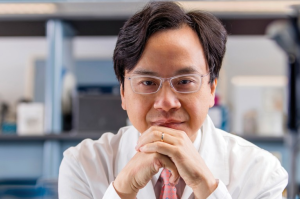 Dennis Lo (left) is the Associate Dean of Research for the Faculty of Medicine at the Chinese University of Hong Kong (China). For the last 25 years, his lab has studied circulating DNA to develop liquid biopsies for prenatal testing, early cancer detection and transplantation monitoring.
Senior Editor Tristan Free caught up with Dennis to discuss fragmentomics and its role in the development of liquid biopsies.
Please explain fragmentomics.
Fragmentomics is a term originally coined in the 1970s that referenced the fragmentation of peptides. However, more recently in our field it has been used to mean the study of the fragmentation of cell-free DNA in blood. Fragmentomics can be used synergistically in liquid biopsies alongside more established genetic markers such as mutations. For example, non-invasive prenatal testing (NIPT) currently uses a combination of DNA fragment count and size. Recent developments in the field have also highlighted the insights that can be gained by investigating the interaction between fragmentomics and epigenetics.
How can fragmentomics be implemented in the development of liquid biopsies?
In the early days of circulating DNA analysis, researchers were primarily interested in finding genetic markers. So, in the past for example, if I was looking at circulating fetal DNA versus a pregnant mother’s DNA, I would try to find a genetic marker that was only present in the baby; let’s say the baby’s a boy, then I would look for Y chromosomal markers. Then let’s say that baby was Rhesus positive, I would look at the Rhesus D gene in the Rhesus-negative mother (who lacks the gene) to enable prenatal fetal blood group genotyping. In cancer, I can look at a variety of cancer-associated mutations. But of course, the problem is that when you start to look at mutations that might cause cancer, the search space is generally huge, making this challenging.
Now, we and others have found that the way DNA fragments into pieces is not random, and it can tell you the tissue of origin for a piece of DNA floating in blood. We have found that the baby’s circulating DNA in the mother’s blood is a bit shorter than the mother’s own DNA. And similarly, the DNA from a cancer is also shorter than that of the non-cancer DNA in blood. So this is the first generation of fragmentomic markers, which are based on size. We have also found that you can actually look at the ends of the fragments to determine which nucleases, enzymes that cut DNA, were involved in creating these fragments, some leave blunt ends, while others generate jagged ends for example. These nucleases may be expressed in different ways in different tissues and in tumor tissues. So that is another type of fragmentomic marker.
 Sowing the seeds of diagnosis: a novel assay for abnormal α-synuclein detection
A new study has developed an assay that can identify incorrectly folded α-synuclein, a biomarker of synucleinopathies, in patient serum.
How have you used fragmentomics to improve current liquid biopsy diagnostics?
If you’re looking at mutations, then only one in many circulating DNA molecules might possibly contain a mutation; it’s the same for methylation patterns. However, in fragmentomics, every cell-free DNA that you find potentially has fragmentomic information that can be used, which is an advantage.
For instance, in the field of non-invasive prenatal testing, previously, the first-generation NIPT was based on counting molecules and trying to look at the relative number of molecules on different chromosomes. For example, an individual with Down syndrome has more molecules on chromosome 21 due to an extra chromosome. Fragmentomic markers can tell you the size distribution of DNA molecules from different chromosomes. So, if a baby has an extra chromosome 21, then the size distribution from chromosome 21 will be a bit shorter than a baby without an extra chromosome.
If there was one thing that you could ask for to improve our understanding of fragmentomics and further our ability to detect and analyze cell-free nucleic acids, what would it be?
The amount of cell-free nucleic acids in various bodily fluids is typically very low, which makes it like searching for gold dust, and then with the protocols that we use nowadays, we are only able to look at a subset of those molecules. So, you really must decide what is most important for your analysis. For example, if you are investigating DNA methylation, then many people will use bisulfite conversion, which may degrade maybe 50% to 90% of DNA to start with; you’re really investing heavily just to look at methylation markers, as it will prevent you from being able to explore other types of nucleic acids due to the massive reduction in molecules available in your sample.
Another instance of this is cell-free RNA. In my lab, we’ve been investigating cell-free RNA since 1999 and we know that RNA is typically less stable than DNA, so we need to use using stabilizing reagents on cell-free RNA. However, these stabilizing agents may not always allow you to conduct an analysis of cell-free DNA, as they are not compatible with the protocols. If I could ask for one thing to improve fragmentomics studies, I would ask for a robust way to look at DNA markers, RNA markers, methylation markers and fragmentomic markers, all in one very small sample.
You were recently presented with two awards for your research. Can you tell us a bit about them?
So recently, I had the honor of being presented with two awards. I received a Breakthrough Prize in Life Science for my work on non-invasive prenatal testing. In September 2022, I also had the honor of receiving the Lasker-DeBakey Clinical Medical Research Award also for this work.