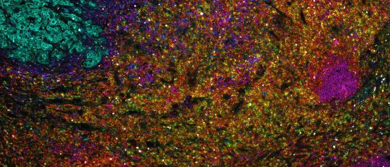
An end-to-end workflow for exploring spatial biology through multiplex IHC

Multiplex immunohistochemistry (IHC) technologies are powerful tools to visualize multiple markers in the same tissue sample. These techniques, which include tyramide signal amplification and cyclic immunofluorescence, help scientists understand the cells that make up a tissue, what markers they express, their spatial distribution and their potential interactions with one another. These methods depend on antibodies that are specific to the target, produce a clean signal and have low background.
In this White Paper, you will find an overview of Fortis Life Sciences workflow for multiplex imaging using cyclic immunofluorescence.
Fortis Life Sciences provide end-to-end custom multiplex imaging and histology services to encompass the full spectrum of the imaging workflow. They also offer pre-formatted panels and application-validated antibodies for multiplex imaging.