VascuViz: a novel method to uncover the secrets of blood vessels

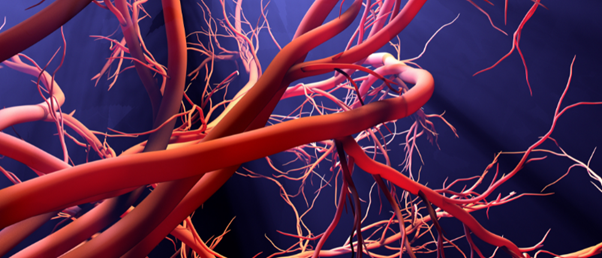
3D images of vasculature systems can now be achieved using a combination of two contrast agents, allowing researchers to perform multiscale imaging of blood vessels.
Researchers at Johns Hopkins Medicine (MD, USA) have developed a novel imaging agent for blood vessels, referred to as ’VascuViz’, which is compatible with several imaging techniques, unlike current more restrictive methods. This uses a quick-setting polymer mixture to fill blood vessels prior to imaging and allows researchers to visualize one sample at different scales.
Often researchers will use techniques such as MRI, CT scans or microscopy to capture images of blood vessels and study them. However, different imaging agents are needed to make blood vessels visible for each of these techniques and can often make them invisible to other imaging methods, presenting difficulties for observing macro- and microvasculature structures simultaneously.
“Usually, if you want to gather data on blood vessels in a given tissue and combine it with all of its surrounding context like the structure and the types of cells growing there, you have to re-label the tissue several times, acquire multiple images and piece together the complementary information,” explains Arvind Pathak, who leads this research group. “This can be an expensive and time-consuming process that risks destroying the tissue’s architecture, precluding our ability to use the combined information in novel ways.”
 Engineered spinal cord implants provide hope for those with traumatic injury
Using fatty tissue, called adipose tissue, researchers have created an implant that restored movement in both acute and chronic models of paralysis.
The research group hopes that VascuViz will accelerate imaging-based research as it enables researchers to collect more data from a single sample by using one imaging agent that is applicable to a variety of techniques.
“Now, rather than using an approximation, we can more precisely estimate features like blood flow in actual blood vessels and combine it with complementary information, such as cell density,” says Akanksha Bhargava, the lead author on this paper.
Bhargava looked at many combinations of imaging agents that are currently used and tested them with different imaging techniques. Bhargava found that combining a CT contrast agent and a fluorescently labeled MRI contrast agent (BriteVu and Galbumin-Rhodamine) would be suitable for several optical-imaging techniques and make the macro- and microvascular structures visible at the same time.
As VascuViz was successful in test tubes, the research group tested it in different mouse tissues, such as the vascular system of breast-cancer models and kidney tissues. 3D visualizations of the vasculature structure of these were created by combining the images collected using MRI and CT scans and optical microscopy. This approach can be combined with mathematical models or images of other tissue elements to understand diseases with abnormal blood flow, such as cancer and stroke.
VascuViz is especially useful for generating computerized visualizations of complex biological systems, for example the circulatory system, and is a new tool in the growing field of “image-based” vascular systems biology. The researchers hope this will improve understanding of the structure of tissue dynamics and their response to drug treatments.