How Pseudomonas aeruginosa penetrates the lungs

Scientists now know how Pseudomonas aeruginosa infects the lungs thanks to human stem cell-derived lung microtissues.
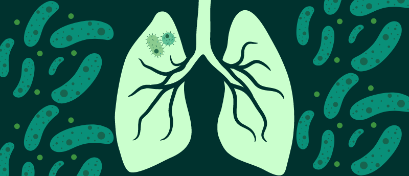
Researchers at the Center for Molecular Life Sciences at the University of Basel (Switzerland) have grown lung microtissues from human stem cells in the lab to uncover how bacteria infect the lungs. Specifically, the team was investigating Pseudomonas aeruginosa (P. aeruginosa), a bacterial pathogen known for causing life-threatening pneumonia. However, until now, scientists didn’t know how this pathogen breaches the protective barriers in the lungs.
P. aeruginosa has developed several strategies to infect humans. Boasting antibiotic resistance and a high mortality rate, P. aeruginosa has made its way onto the WHO’s list of the 12 most dangerous bacterial pathogens. It can breach the lungs’ thin, mucus-covered layer of tightly packed cells, which protects the deeper layers of lung tissue. The mucus layer traps other pathogens and prevents infection; however, this is not the case for P. aeruginosa.
To investigate the mechanism of infection, the researchers grew stem cell-derived lung microtissues. “We have grown human lung microtissues that realistically mimic the infection process inside a patient’s body,” explained senior author Urs Jenal. “These lung models enabled us to uncover the pathogen’s infection strategy. It uses the mucus-producing goblet cells as Trojan horses to invade and cross the barrier tissue. By targeting the goblet cells, which make up only a small part of the lung mucosa, the bacteria can breach the defense line and open the gate.”

Coming of phage: treating antibiotic-resistant infections with bacteriophages
In this interview, Martha Clokie talks about phages’ diverse structure and function, the roles that both natural and engineered phages could play in overcoming the growing issue of antibiotic-resistant bacterial infections and what happens if bacteria become phage-resistant.
The pathogen’s ability to hijack the mucus-producing goblet cells, replicate within them and kill them – bursting the cell and creating ruptures in the protective barrier – is what allows them to infect the lungs. At the points of rupture, the bacteria colonize, taking advantage of the vulnerable tissue to spread deeper into the lungs.
In addition to illuminating the infection strategy of P. aeruginosa, this research initiative has also shed light on how the pathogen changes its behavior throughout the course of infection, another aspect that had eluded scientists for many years. Thanks to a small signaling molecule called c-di-GMP, we know that the pathogen does rapidly change its behavior. Now, the team has developed a biosensor that can track c-di-GMP in individual bacteria.
“This is a technological breakthrough,” commented Jenal. “Now we can monitor in real time and with high resolution how this signaling molecule is regulated during infection and how it controls the pathogen’s virulence. We now have a detailed view on when and where individual bacterial cells activate certain programs to regulate their behavior. This method enables us to investigate lung infections in more detail.”
Not only have these lung organoids provided insights into the infection process of P. aeruginosa, but they also offer researchers the ability to study the effects of antibiotics in tissue. Human tissue organoids have already transformed our understanding of human disease; they will continue to be essential to our development of effective strategies to combat pathogens.