The effect of temperature fluctuations on the evolving coronavirus: speculations by Morteza Mahmoudi

Temperature has long been recognized as a major determinant of spontaneous mutation and as playing a critical role in the mutation rate of bacteria and viruses [1–6]. Reasons behind this phenomenon include the existence of temperature-sensitive mutants in a wide range of viruses, including the influenza A virus [7, 8].
Some species, like bats, are natural reservoirs of viruses. As some bats such as horseshoe bats and noctules have no mechanisms for regulating temperature [9], their viral reservoirs may be subject to constant temperature variation (~17–35°C). It was recently found that environmental fluctuations have the capacity to significantly alter the fate of mutations [9]. Therefore, it is reasonable to speculate that viruses that use bats as reservoirs may be even more prone to dramatic and less predictable mutations.
Studies show that climate change can significantly influence a bat’s habits and life cycle, including the timing of hibernation [10–13]. These alterations may also affect the temperature fluctuation within the bodies of bats, which may in turn produce unexpected mutations in the viruses for which bats serve as natural reservoirs. The current outbreak of COVID-19, an unprecedented mutation of a coronavirus, may provide evidence to support this speculation, as bats are likely to be the main reservoir of coronaviruses [14–16].
Sign up to The Nanomed Zone for free
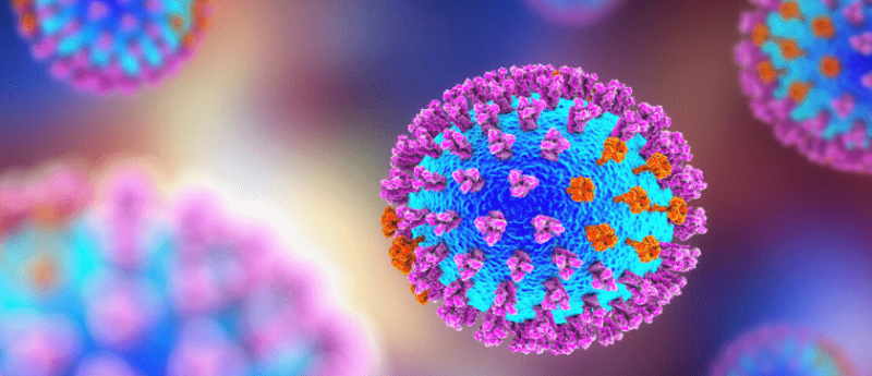
We have studied the role of slight temperature variations in the functionality and affinity properties of proteins (in terms of their interactions with nanoparticles) and found that even slight temperature changes (e.g., a few centigrade degrees centigrade) may affect some protein characteristics [17–19]. We used both experimental (e.g., by fluorescence correlation spectroscopy) and computational (e.g., molecular dynamics) approaches to show that the slight temperature changes can change the binding site of proteins with nanoparticles and therefore change the biological identity of nanoparticles (i.e., a layer of biomolecules that covers the surface of nanoparticles [20, 21], which is like spike proteins on the surface of coronavirus), which affects their cellular uptake and toxicities [17, 19, 22]. Therefore, one may also speculate that temperature variations in the viral host might alter the functionality and affinity of spike proteins (specifically on their unstructured sections) on the surface of coronaviruses. Deeper understanding of the part of spike proteins that can be affected by temperature variations can help researchers to better understand other possible binding sites of spike proteins to human receptors (and/or their stickiness to the cell membrane), which helps scientists to design complementary drugs/approaches to minimize virus resilience to specific therapeutic approaches. Electron tomography and 3D cryo-TEM approaches in combination with image analysis and computational approaches can also shed more light on the 3D structure of virus spike proteins and their possible dependency on temperature variations.
As extensive multi-disciplinary research studies [23–26] (and even those within nanomedicine itself [27, 28]) are addressing the COVID-19 pandemic, the scientific community should also consider the crucial effects of temperature, not only for prediction of possible future mutations of coronaviruses, but also with regard to proposed diagnostic and therapeutic approaches.
Among the hard lessons we are gradually learning from this emerging pandemic is the price to be paid for the lack of proactive integration among all stakeholders. It is noteworthy that with COVID-19, the stakeholders encompass almost all the “humanity” across nationalities, cultures and expertise. I believe we must all learn to be much more proactive than reactive regarding such events. Although specific scientific communities (e.g., virology) may play central roles in helping to control this pandemic, I believe that the most effective resolution to this situation and similar future challenges is integrated functioning of all stakeholders; this includes scientists (regardless of their area of expertise), funding agencies, legislators/governors, fast-track commercialization experts and the general public. Such proactive and integrated functioning among all stakeholders would constitute a unique and ready-to-use platform for response to future pandemics. For COVID-19, however, such collaboration is just beginning to coalesce. For example, funding agencies are now joining the fight against COVID-19 through i) announcing new, rapid funding to support COVID-19 research studies (e.g., American Heart Association [29]) and ii) allowing scientists to shift their existing/renewal grants for use in COVID-19-related research (e.g., National Institutes of Health [30]).
The COVID-19 outbreak is showing us that we are much more vulnerable than we thought to such unexpected mutations in viruses. The prediction of any future mutations, more proactive action responding to the growing issue of climate change and other factors that may affect the mutation of the coronavirus, together with the creation of a comprehensive unified global platform for rapid response to pandemic crisis (through integrated functioning of all stakeholders), may help us to address future outbreaks more adequately and in a more timely manner, saving more lives and significantly reducing the social and economic burdens posed by such disasters.
The opinions expressed in this feature are those of the author and do not necessarily reflect the views of The Nanomed Zone or Future Science Group.
For more free COVID-19 related content please visit our dedicated COVID-19 Hub on Infectious Diseases Hub here.
For more related news from The Nanomed Zone be sure to sign up here.