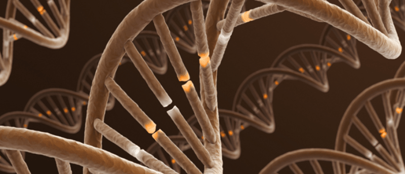
Origami DNA is unfolding the mysteries of mechanical cell signaling

A three-dimensional DNA origami robot has been developed to probe the mechanism of recently discovered cellular receptors sensitive to touch and force.
On a cellular level, our bodies have a dense network of signaling pathways and cellular processes that rely on a constant transmission of signals. Chemical and electrical signaling mechanisms are the predominant pathways; however, they are not the only methods through which our cells send invisible messages.
The recent Nobel prize-winning discovery of cellular mechanoreceptors, receptors that are triggered to produce chemical signals when a physical force is applied to them, has opened a new frontier to further our understanding of the macromolecular world. These receptors are vital regulators of processes such as vasoconstriction and the perception of pain and sound. They even play a role in our ability to breathe.
A coalition of researchers from the National Institute of Health and Medical Research (INSERM; Paris, France), National Center for Scientific Research (CNRS; Paris, France) and the University of Montpellier (France) have developed a DNA origami self-assembly nanostructure to probe the mechanism of these receptors. DNA origami is the controlled folding of nucleic acids to create dynamic two- and three-dimensional structures and is a nanotechnological technique that has advanced the field significantly over the last decade.
 Regulatory mechanism for centromere distribution uncovered
Researchers propose a two-step regulatory mechanism for non-Rabl configurations of centromeres, which are chromosomal domains linking pairs of sister chromatids.
For this study, the team exploited the technique to develop a ‘nano-robot’ (a microscopic winch) with one purpose, to apply a trillionth of a newton (piconewton) of force to mechanoreceptors. To put this in perspective, the force due to gravity exerted on the apple that dangled over the eponymous Isaac Newton’s head one fateful afternoon was approximately one newton.
The robots were coupled with a molecule designed for recognition of the mechanoreceptor. With these robots, researchers will be able to further probe the mechanisms of mechanosensitive receptors and potentially find other receptors that are sensitive to force and are yet to be discovered. Additionally, insight into the precise moment an applied force activates cascading cellular pathways will allow scientists to expand our understanding of the importance of these stimuli.
This study also marked the first recorded instance of a synthesized, self-assembled DNA object capable of applying forces on a piconewton scale accurately.
Looking forward, the team is confident that this is a major landmark of nanotechnology, with myriad future applications, yet is still very much in its infancy. “The design of a robot enabling the in vitro and in vivo application of piconewton forces meets a growing demand in the scientific community and represents a major technological advance,” explained senior author Gaëtan Bellot (INSERM). “However, the biocompatibility of the robot can be considered both an advantage for in vivo applications but may also represent a weakness with sensitivity to enzymes that can degrade DNA. So, our next step will be to study how we can modify the surface of the robot so that it is less sensitive to the action of enzymes”.