Finding our way to an atlas of the human brain

Researchers have developed technology that can characterize different populations of human brain cells, a critical step toward the reconstruction of a 3D brain model.
A group of investigators from Massachusetts General Hospital (MGH; MA, USA) has recently constructed a cellular atlas of a region of the human brain. Using a combination of innovative imaging techniques, the researchers characterized different brain cell types and their distribution in Broca’s area, a region of the brain associated with speech. The technology developed in this study could enable the reconstruction of a highly accurate and detailed 3D model of the human brain; such a model is critical to future study of brain function and disorder.
The human brain is an incredibly complex organ made up of billions of cells, all organized in complex three-dimensional structures. To understand the brain’s function, it’s important to characterize its cellular architecture.
Despite recent advances in imaging technologies, no current technology can visualize the microscopic details of the brain without significant distortion. This distortion is often a result of the techniques used in histological imaging. Until now, researchers have struggled to produce images of a high enough resolution to be able to build an accurate model of the human brain.
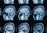
In the study, published in Science Advances, the team at Massachusetts General Hospital looked to overcome the limitations associated with singular imaging techniques. They did this by bridging data from macroscopic magnetic resonance imaging (MRI) and microscopic light-sheet fluorescence microscopy (LSFM) using a mesoscopic imaging technique called optimal coherence tomography (OCT).
In a human postmortem specimen, the researchers performed an initial MRI to image the whole brain and provide a reference coordinate system for the cellular atlas. They then proceeded to image Broca’s area, specifically, using serial-sectioning OCT.
 Mapping the brain: from mice to mankind
A collaborative projects attempts to chart every neural connection in the mouse brain and create a comprehensive connectome.
OCT provides high-resolution images of cross-sections of the brain up to several hundred micrometers in depth. The integration of a mechanical sectioning device enabled further imaging of tissue sections up to centimeters in each dimension. By performing imaging before sectioning, the complex three-dimensional information was preserved, which facilitated the more accurate registration of data to the MRI-based coordinate system.
Using confocal LSFM and an advanced tissue transformation protocol called SHORT, the researchers then characterized sections to the microscopic level, labeling, quantifying and visualizing the different brain cell populations. This cellular information was then integrated into the whole-brain reference.
The result of this study was a micrometer-resolution reconstruction of the cellular architecture of this specific region of the human brain.
“We built the technology needed to integrate information across many orders of magnitude in spatial scale from images in which pixels are a few microns to those that image the entire brain,” explained co-senior author Bruce Fischl (MGH).
“These advances will help us understand the mesoscopic structure of the human brain that we know little about. Structures that are too large and geometrically complicated to be analyzed by looking at 2D slices on the stage of a standard microscope, but too small to see routinely in living human brains.”
The innovative technology developed in this study could be extended to reconstruct a three-dimensional cellular model of the whole brain, an important tool in the investigation of intra- and inter-individual brain cell variations. Most importantly, such a model could offer a unique insight into the pathological changes that happen in neurodegenerative and psychiatric illnesses; for example, the identification of brain cells that are affected by illness, or the heterogeneity within cell types and their spatial distribution in brain disorders.
“Currently we don’t have rigorous normative standards for brain structure at this spatial scale, making it difficult to quantify the effects of disorders that may impact it such as epilepsy, autism, and Alzheimer’s disease,” stated Fischl.
