Largest brain wiring diagram generated from grain-sized sample
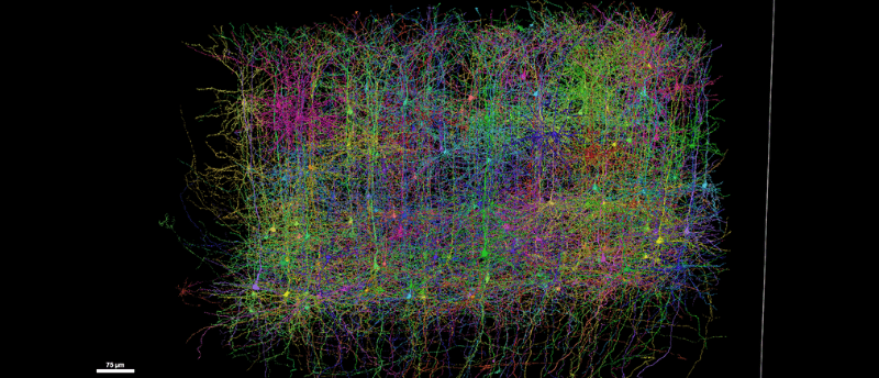
A global team has constructed the most detailed wiring diagram of a mammalian brain to date.
A global collaboration of over 150 researchers has created the most detailed wiring diagram of a mammalian brain to date, mapping the complete structure and function of a grain-sized piece of mouse visual cortex. The Machine Intelligence from Cortical Networks (MICrONS) project aims to understand how neural circuits give rise to perception and cognition.
In 1979, famed molecular biologist Francis Crick wrote, “It is no use asking for the impossible, such as, say, the exact wiring diagram for a cubic millimeter of brain tissue and the way all its neurons are firing.” Since then, technology has developed so that the once impossible is now a step closer to reality.
To create the map, researchers at Baylor College of Medicine (TX, USA) performed two-photon in vivo calcium imaging on around 75,000 neurons in the primary visual cortex and higher visual areas of a mouse brain while it watched different movies and YouTube videos.

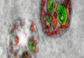
How do brain cancers reprogram immune cells to encourage tumor growth?
A mechanism underlying neutrophil reprogramming in brain cancers has been uncovered, illuminating potential therapeutic targets to stop their pro-cancer effect.
The mouse was then shipped to the Allen Institute (WA, USA), where they extracted the imaged tissue, sliced it into more than 25,000 layers and imaged the slices using electron microscopy. Researchers from Princeton University (NJ, USA) then used AI and machine learning to reconstruct the cells and connections into a 3D volume. The final dataset includes over 200,000 cells, 4 km of axons and 523 million synapses, and is freely available through the MICrONS Explorer.
The map was published alongside studies that reveal new cell types, characteristics, organizational and functional principles, and a new way to classify cells. This included the discovery that inhibitory neurons are far more selective and organized than assumed. Some inhibitory neurons were found to cooperate to suppress multiple excitatory cells, while others were more precise, targeting specific cells.
Beyond mapping healthy brain circuits, the project may also provide a blueprint for studying neurological conditions. “If you have a broken radio and you have the circuit diagram, you’ll be in a better position to fix it,” said Nuno da Costa (Allen Institute), a member of the MICrONS consortium. “We are describing a kind of Google map or blueprint of this grain of sand. In the future, we can use this to compare the brain wiring in a healthy mouse to the brain wiring in a model of disease.”
The MICrONS team emphasizes that this work lays the foundation for larger-scale efforts. “IARPA’s moonshot investment in the MICrONS program has shattered previous technological limitations, creating the first platform to study the relationship between neural structure and function at scales necessary to understand intelligence,” commented David A. Markowitz, former Intelligence Advanced Research Projects Activity (IARPA) program manager who coordinated this work. “This achievement validates our focused research approach and sets the stage for future scaling to the whole brain level.”