Mapping the larval fruit fly connectome

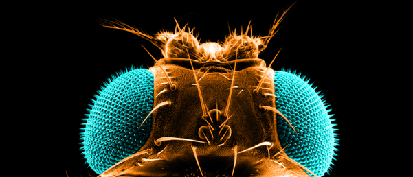
A new study has completely mapped the larval fruit fly brain for the first time, illuminating the positions of 3,016 neurons and how they connect.
An international team led by researchers from the University of Cambridge (UK) and Johns Hopkins University (MD, USA) have successfully built a comprehensive map of the larval Drosophila melanogaster’s brain. This positional information and patterns of connectivity provide a basis upon which researchers can build a mechanistic understanding of brain function.
Compared to previous brain mapping studies that have succeeded in mapping the roundworm brain, the current study has accomplished the mapping of a more complex neuronal system. The larval fruit fly brain is similar to its more senior counterpart as well as other large insects; additionally, the larval fruit fly has a sophisticated behavioral range, including action-selection and learning, which makes them attractive organisms for brain mapping.
The development of electron microscopy has revolutionized researchers’ ability to image whole brains; the current study utilized this technology to reconstruct the neural circuitry of the larval fruit fly brain. The team imaged thousands of slices of a larval fruit fly brain, annotating the neuronal connections and mapping 3,016 neurons and 548,000 synapses.
 Studying honey bee brains in vivo
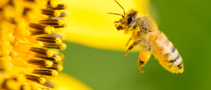
Researchers integrate a fluorescent calcium sensor into honey bee brains to monitor the areas that are activated in response to environmental stimuli.
They also developed computational tools to recognize circuit patterns and likely information pathways using a deep learning architecture. “The most challenging aspect of this work was understanding and interpreting what we saw. We were faced with a complex neural circuit with lots of structure. In collaboration with Professor Priebe and Professor Vogestein’s [paper co-authors] groups at Johns Hopkins University, we developed computational tools to predict the relevant behaviors from the structures. By comparing this biological system, we can potentially also inspire better artificial networks,” explained senior author Marta Zlatic (University of Cambridge).
Although technologies have advanced whole-brain imaging, they aren’t yet powerful enough to map specific neurons and their connections in more complex organisms; however, the larval fruit fly connectome provides an important step in the right direction, serving as a reference for future brain mapping studies.
“All brains of all species have to perform many complex behaviors: for example, they all need to process sensory information, learn, choose food, and navigate their environment. In the same way that genes are conserved across the animal kingdom, I think that the basic circuit patterns that drive these fundamental behaviors will also be conserved,” commented Zlatic.
In the future, the researchers plan to investigate the brain circuitry associated with specific behaviors, taking the connectome one step further to match behavior with its physical position in the brain.


