Gearing up for replication
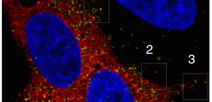
A new technique allows researchers to simultaneously monitor RNA, DNA, and protein trafficking during HIV’s replication cycle.
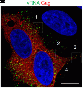
Immunofluorescent visualization of viral RNA and viral proteins in virions (1).
HIV’s integration into a cell functions similar to a network of little gears moving a larger gear, the host cell. The little gears are the viral components of HIV (DNA, RNA, and protein) that facilitate infection. Current HIV treatments are designed to target viral replication. However, despite significant technical progress in individually visualizing the movements of the nucleic acids and proteins carrying out viral infection, researchers have been unable to simultaneously track how all of these gears move in concert to successfully infect a cell.
“You want to see transcription from an integration site, and so far, this has not been possible in the case of HIV,” said Stefan Sarafianos from Emory University. Now, in the journal Nature Communications, Sarafianos and his colleagues reveal a novel microscopy technique that allows researchers to dynamically and temporally track changes in viral RNA, DNA, and protein at a single-cell resolution (1). This integrated approach, called multiplex immunofluorescent cell-based detection of DNA, RNA, and Protein (MICDDRP), allowed researchers to track HIV integration across the replication cycle for the first time.
Using a technique akin to fluorescent in situ hybridization (FISH), the researchers designed specific probes to label nucleic acid intermediates during HIV infection, a technique that is highly compatible with protein immunofluorescence. This gave researchers a peak into how the gears in the system start moving during early infection. For example, by specifically labeling the first strand of synthesized viral DNA, the researchers found that reverse transcription starts in the cytoplasm.
Furthermore, consistent with predominant models of HIV replication, the probes detected a reduction in viral RNA and an increase in viral DNA during reverse transcription prior to viral DNA being shuttled to the nucleus. “We could really follow the kinetics of reverse transcription – how DNA is made while RNA is being degraded through the RNase activity,” said Sarafianos.
Interestingly, the team went on to show that after reverse transcription there was a transcriptional burst, or an accumulation of nascent viral RNA in the nucleus that then travels back to the cytoplasm. “Transcriptional bursts have been reported in transcriptional cellular genes but we now show them in HIV and an infectious system,” said Sarafianos.
Understanding HIV’s replication cycle may guide the development of antiviral therapies designed to inhibit the gears in the system from working properly. Furthermore, Sarafianos and colleagues hope that this technique could be applied to evaluate and asses the efficacy of drugs that reactivate HIV from latency.


