The usual suspect: solving the case of RNase contamination in RNA analysis
RNase contamination can cause chaos in RNA analysis labs. What are these problems and how can they be solved?
RNase: the usual suspect
RNase is the enzyme responsible for RNA degradation in living organisms. They play a key role in the maturation of RNA molecules and are a first line of defense against RNA-containing viruses, as well as in the break-down of old RNA. There are many different types of RNase, with the main type utilized in labs being RNase A, which specifically targets single-stranded RNA.
RNase A is commonly described as one of the hardiest enzymes in lab usage. RNases are generally very rich in disulfide bonds, which make them incredibly stable enzymes and can even survive autoclaving.
Whilst RNase is a necessary component of many biological processes, it is best described as a pain and a hindrance to those working in RNA analysis.
It is incredibly easy to contaminate a RNA sequencing lab with RNase, yet notoriously difficult to ensure complete eradication. This is due to the presence of environmental RNase in microbial sources in the air as well as from human skin, hair or saliva.
Contamination: the crime
In order for RNA experiments to be successful, it seems obvious that RNA molecules must be as intact as possible. However, due to the interference of RNase, which quickly breaks down RNA molecules when present, this is often not the case. Researchers can then have abnormalities thrown at them, inconsistent results, which cause frustration and confusion.
“RNases are pervasive and when contaminating critical surfaces can present problems for laboratories engaged in RNA sequencing and analysis. Miniscule amounts of common enzymes, such as RNase, can make an experimental series inaccurate. Standard techniques for decontaminating laboratory equipment can take hours,” commented Theresa Thompson, an application scientist at Phoseon Technology (OR, USA).
Preventing the physical presence of RNase in a ‘normal’ lab would be an unrealistic expectation. Instead, managing the situation thoroughly is a more realistic aspiration. Researchers can keep RNase contaminations to a minimum by frequently opening fresh disposables and using new pairs of gloves; a situation which may seem all too familiar to the ever careful RNA analyst. On top of this, it is, however, possible to inactivate RNase molecules by various methods, halting their activity and rendering them useless against RNA.
These current methods of RNase inactivation tend to be costly, time-consuming and wasteful. They include DEPC treatment of water and autoclaving, chemical decontamination of surfaces and chemical treatment of equipment followed by rinsing in RNase-free water and baking glassware.
It is difficult to know when a lab is clean enough. All it could take is to leave a pipette on a desk to pick up RNase from shed skin cells or begin to use an autoclaved piece of equipment only for it to immediately begin to infer new RNase molecules from the air. Autoclaving also does not destroy all RNase activity on its own, as they can retain partial activity on cooling to room temperature.
Whilst chemical cleaning may deactivate the enzyme, it leaves behind chemical residues that can potentially contaminate the experiment in other ways.
There is an emerging method of RNase decontamination that has shown promising results and is quick and chemical-free. UV light can irreversibly inactivate RNase, with studies demonstrating this is possible in less than 1 minute.
Let there be light!
Phoseon Technology has developed the use of UV light for RNase decontamination into a novel technology:
“KeyPro™ Technology is a high-intensity UV LED microplate decontamination system. An array of diodes puts out high-intensity UV light in two wavelengths – 275 nm and 365 nm – proven to rapidly and reliably inactivate RNase. As the array scans across the decontamination chamber, the light reaches all top-exposed areas of the slide or plate,” explained Thompson.
“Complete inactivation of laboratory contaminants, including RNase A, can be accomplished by the KeyPro system in under 5 minutes and at a fraction of the cost of traditional methods.”
The KeyPro Technology is currently targeted at microplate, preparatory slide and small surface decontamination and works by a scanning array of UV LEDs in combination with an adjustable height platform.
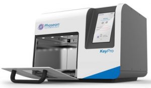
The KeyPro system.
The use of UV light eliminates much of the issues associated with the current methods of decontamination. There is no need to repeatedly rinse equipment afterwards with RNase-free water and there is no chemical residue, which cuts down time and potential contamination through other routes. There is also a lesser need for disposables, reducing cost and waste.
Studies have also shown that all common laboratory surfaces, including plastic, can be safely decontaminated through this use of UV light.
It’s been shown that the protocols, which can benefit from this include multiple types of RNA sequencing, ribosome profiling and next generation sequencing of RNAs.
It is hoped this technology can be further developed to enable decontamination of bigger pieces of equipment as well as manipulation of light wavelengths, as Thompson concluded:
“A model with a larger chamber is planned for more flexibility in what equipment can be decontaminated. Beyond that, we have found that other wavelengths of light can be used to manipulate biomolecule function, such as accelerate enzyme activity or selectively denature only certain parts of the molecular structure of a protein.”
