A peek behind the paper: 3D mapping and accelerated super-resolution imaging of the human genome using in situ sequencing

Take a look behind the scenes of an article recently published in Nature Methods entitled ‘3D mapping and accelerated super-resolution imaging of the human genome using in situ sequencing’, as we invite authors, Huy Nguyen, Shyamtanu Chattoraj and David Castillo, to shed some light on their recent research.
Please can you give us a short summary of the methods presented in your Nature Methods article, “3D mapping and accelerated super-resolution imaging of the human genome using in situ sequencing”?
In our recent Nature Methods paper, we simultaneously imaged up to 249 single-copy genomic regions across all 46 chromosomes inside a single cell nucleus derived from human skin. This gave us a genome-wide view of human chromosomes and individual genomic regions at a scale that, at the time we published, had not been seen before. We designed OligoFISSEQ as a versatile technology that allows its use in many different scenarios, such as in combination with existing super-resolution methods, drastically reducing the time required for obtaining nanoscale images of the genome. OligoFISSEQ is also compatible with immunofluorescence, enabling the simultaneous imaging of proteins and DNA.
Like this? Get in touch to publish your own Peek Behind the Paper now!
What scientific problem inspired you to develop OligoFISSEQ?
The human genome has been sequenced (although not fully) since 2003 and still, there remain more questions than answers as to how DNA impacts gene expression and ultimately, health and disease. Furthermore, not only is the genome very complex, it is folded and compacted inside a tiny nucleus. To understand how the genome functions and regulates several important biological processes, it’s necessary to visualize many genomic regions simultaneously, not just a few, hence the reason for developing OligoFISSEQ. For example, we know that there is high structural variability of specific genomic features from cell to cell, but we still don’t know if those local changes have a global impact in the organization of the whole genome. Does a local change in one chromosome induce a change in the neighboring chromosome? And in the neighbor of the neighbor? It might be that what we think of as local variability is part of a global regulatory mechanism to contribute to the core machinery of the cell.
Of note, in many diseases including cancer, there are a lot of chromosomal aberrations, wherein certain chromosomes change their location inside the nucleus. In addition, parts of chromosomes can become embedded in other chromosomes. Oftentimes, these are signatures of the onset of disease. So, we need to see all the chromosomes at fine scale resolution in order to understand such phenomena. This was one of the main inspirations to develop OligoFISSEQ. The more you see, the more you believe!
What impact do you hope OligoFISSEQ will have in genomics, and for researchers working in the field?
What we provide to the community is a spatial genomics technology that makes possible the visualization of hundreds of regions of interest at the same time in individual cells at reasonable cost and in a time efficient manner. Ultimately, OligoFISSEQ should enhance the potential of genome research many fold! It enables researchers to study the structure of the genome in an accelerated and cost-effective way and with enough statistical power to be able to discern if certain chromosome conformations reflect cell regulation or randomness. This information will provide further insight into how the genome operates and whether loci operate individually or in a coordinated manner. We like to describe OligoFISSEQ as a ‘one stop shopping market for genome visualization’.
How does OligoFISSEQ compare to other methods – in terms of cost, ease of use, speed and so on?
OligoFISSEQ is actually a suite of three in situ sequencing technologies that enable researchers to decode barcodes embedded into an initial library of Oligopaint oligos targeting the genome. One method effects sequencing via ligation (SBL), another enables sequencing by synthesis (SBS), and a third uses sequencing by hybridization (SBH). Comparable methods rely solely on SBH. Of the three OligoFISSEQ methods, SBL and SBS are, by far, the more scalable. This is because the cost of hybridization-based methods for decoding barcodes increases with the number of targets being identified, while the cost of decoding by SBL or SBS remain the same whether a researcher is interrogating 1 or thousands of targets. Regarding speed, one could theoretically image tens of thousands of targets at a 50 kilobase genomic resolution, in just 3-4 days. That is where we are aiming for.
Are there any challenges that still remain with this technique that users should look out for?
One of the main challenges that we are still working on is how to image very small genomic regions. Areas that will help us is to improve the ability to image smaller targets are the optics of our microscope and the chemistry of our sequencing. Another challenge is to improve our detection pipeline. We aim to do this by incorporating cutting-edge image processing tools and algorithms. By reducing target size and increasing detection efficiency, we will better be able to map individual genes in healthy and diseased cells.
How complicated is this method to set up, and what equipment would a researcher looking to perform it need to have to hand?
We developed OligoFISSEQ in such a way that it can be incorporated into laboratories that have a basic, diffraction-limited, fluorescence microscope. And, for laboratories that conduct super-resolution microscopy, OligoFISSEQ has the potential to greatly accelerate this level of imaging. We are currently combining OligoFISSEQ with commercial microfluidic systems to further automate imaging and sequencing, but this is not essential to the method.
This was quite a collaborative effort – as a group, what are your top tips for successful collaborations?
This was indeed a very fun collaboration across the globe. Here are some reasons why our collaboration worked so well:
- Our laboratory was in constant communication with other researchers, meeting frequently (in our case it was mostly virtual). This level of communication was critical, because our team was located in both Boston and Barcelona. Our emails were long and frequent, and our virtual meetings occurred nearly around the clock.
- We understood each other’s interests and contributions and truly cared about each other’s welfare. This made it easy, even enjoyable, to resolve differences of opinion.
- We shared comparable levels of passion and interest in the project.
Luckily for us, everyone on the project was highly motivated, and we all got along extremely well.
What are you hoping to do next in this area?
The next steps are to further refine OligoFISSEQ and increase its capacity to image smaller targets in a wide range of biological samples, including other cell types, tissues, organoids, biopsies, and so on. Furthermore, we plan to really scale up the number of targets that can be imaged simultaneously, allowing us to perform single-cell studies involving thousands of cells in the area of structural genomics. We are especially excited about being able to synergize with existing techniques, such as Hi-C, and hope that, with its genome-wide capacity, OligoFISSEQ will enhance our field’s ability to understand the relationship between 3D genome organization and genome function. There are an endless number of questions to be explored!
PUBLISH YOUR OWN PEEK BEHIND THE PAPER NOW
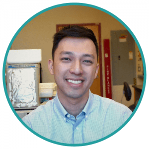
 Huy Nguyen, PhD, completed his genetics PhD work in the laboratory of Giovanni Bosco, from Department of Genetics, Geisel School of Medicine, Dartmouth College (NH, USA). He studied condensins (molecular machines that are involved in organizing chromosomes) in Drosophila and contributed to the discovery of condensin regulators. Nguyen is now a postdoc in the laboratory of Ting Wu, Genetics Department, Harvard Medical School, Harvard University (MA, USA). He is involved in developing technologies for genome visualization and is also interested in how the genome responds to extreme stresses; for example, desiccation, space, radiation.
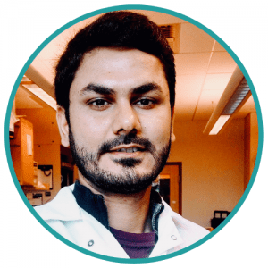
 Shyamtanu Chattoraj, PhD, completed his PhD in 2016 in single molecule biophysics and chemistry at the Indian Association for the Cultivation of Science (Kolkata, India) under the supervision of Kankan Bhattacharyya. During his PhD, Chattoraj developed novel probes for live cell imaging and to understand the physical chemistry of a live cell. Currently he is a senior postdoc in Wu’s lab. He is developing multiplex genome imaging technologies that would enable the visualization of a genome at unprecedented resolution both at diffraction limited as well as super-resolution level.
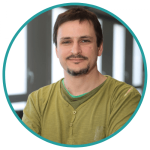
 David Castillo obtained his MSc in photonics from the Universitat Politècnica de Catalunya (Barcelona, Spain) where he worked in super-resolution microscopy. He has a background in physics and engineering. He works as a bioinformatics technician in the structural genomics team of Marc A. Martí-Renom at CNAG-CRG (Barcelona), developing tools for the analysis, modeling and visualization of HiC data. He is also interested in the integration of microscopy to the modeling of genomic 3D structures.